- Therapeutic Cataract & Refractive
- Lens Technology
- Glasses
- Ptosis
- Comprehensive Eye Exams
- AMD
- COVID-19
- DME
- Ocular Surface Disease
- Optic Relief
- Geographic Atrophy
- Cornea
- Conjunctivitis
- LASIK
- Myopia
- Presbyopia
- Allergy
- Nutrition
- Pediatrics
- Retina
- Cataract
- Contact Lenses
- Lid and Lash
- Dry Eye
- Glaucoma
- Refractive Surgery
- Comanagement
- Blepharitis
- OCT
- Patient Care
- Diabetic Eye Disease
- Technology
Case study: persistent keratitis OS presents in 68-year-old woman
A 68-year-old White woman was referred for persistent keratitis OS and failure on multiple medications. Find out her official diagnosis and treatment plan.


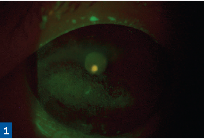
Figure 1. Infectious keratitis before application of Prokera Slim.
Presentation
A 68-year-old White woman was referred for persistent keratitis OS and failure on multiple medications: Restasis, Xiidra, prednisolone 1%, Lotemax 0.5%, and Cequa. She was using Systane preservative-free drops, 4 to 6 times daily. Sodium fluorescein staining revealed marked keratitis OS (Figure 1).


Caldwell

Treatment
I educated the patient about her condition, and after lengthy discussion it was decided that a Prokera Slim amniotic membrane would be used to treat the OS.
Follow-up
I instructed my patient to make an appointment to have the Prokera ring removed when her vision cleared (due to membrane dissolving). Her vision cleared 6 days after Prokera Slim insertion. At this point, she was only using Systane preservative-free artificial tears. The ring was removed, and the patient was placed on autologous serum eye drops 20%, 1 drop 6 times per day.

")
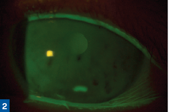
Figure 2. The eye 6 days after application of Prokera Slim. (All figures courtesy of Greg Caldwell, OD, FAAO)
Outcome and observations
The patient presented with a chronic and persistent keratitis OS. The cause was multifactorial with 1 component being toxic keratitis from the various pharmaceuticals used. This patient needed a therapy that supported wound healing and not wound covering. A Prokera Slim was used to provide wound healing. The patient reported no problems or discomfort with Prokera Slim during the 6-day treatment (Figure 2).
The patient responded wonderfully to the therapy and reported increased comfort and improved vision.
READ Dr.Caldwell's full article: Cryopreserved amniotic membrane excels at healing ocular surface wounds