- Therapeutic Cataract & Refractive
- Lens Technology
- Glasses
- Ptosis
- AMD
- COVID-19
- DME
- Ocular Surface Disease
- Optic Relief
- Geographic Atrophy
- Cornea
- Conjunctivitis
- LASIK
- Myopia
- Presbyopia
- Allergy
- Nutrition
- Pediatrics
- Retina
- Cataract
- Contact Lenses
- Lid and Lash
- Dry Eye
- Glaucoma
- Refractive Surgery
- Comanagement
- Blepharitis
- OCT
- Patient Care
- Diabetic Eye Disease
- Technology
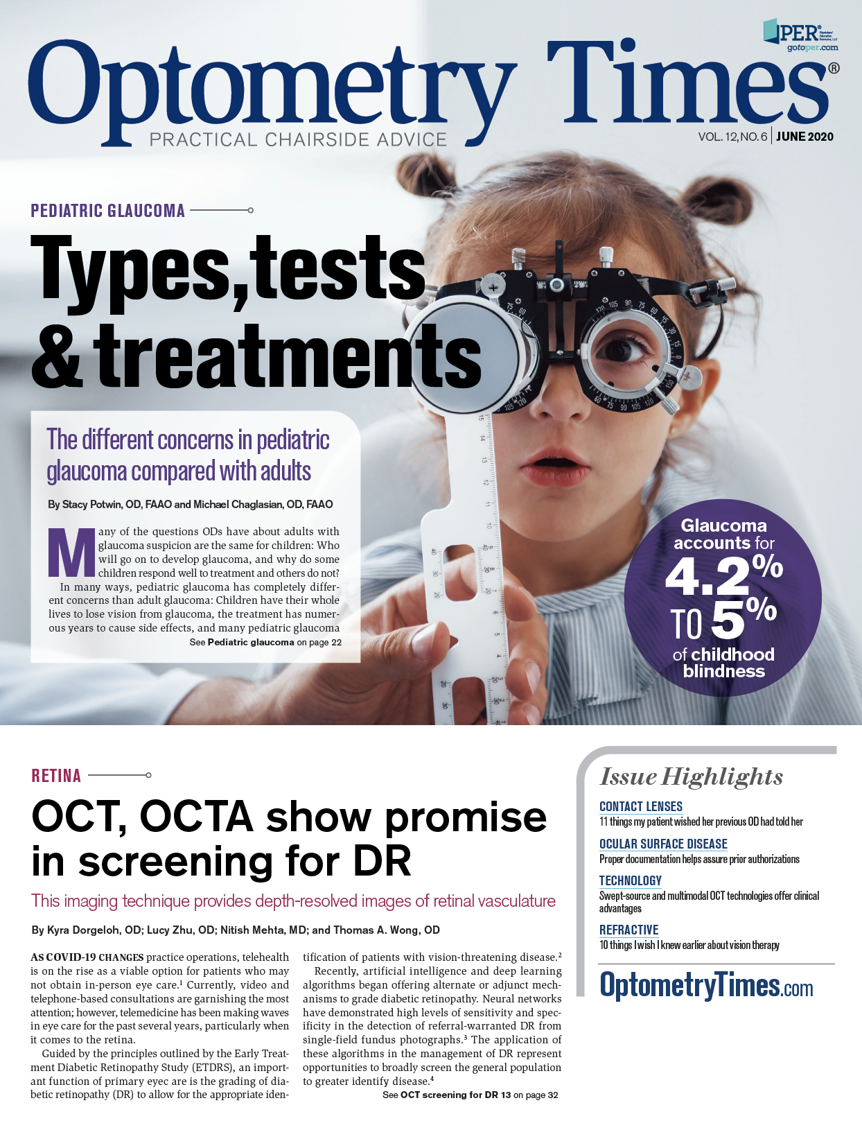
OCT, OCTA show promise in screening for DR
This imaging technique provides depth-resolved images of retinal vasculature.


As COVID-19 changes practice operations, telehealth is on the rise as a viable option for patients who may not obtain in-person eye care.1 Currently, video and telephone-based consultations are garnishing the most attention; however, telemedicine has been making waves in eye care for the past several years, particularly when it comes to the retina.
Guided by the principles outlined by the Early Treatment Diabetic Retinopathy Study (ETDRS), an important function of primary eyec are is the grading of diabetic retinopathy (DR) to allow for the appropriate identification of patients with vision-threatening disease.2
Recently, artificial intelligence and deep learning algorithms began offering alternate or adjunct mechanisms to grade diabetic retinopathy. Neural networks have demonstrated high levels of sensitivity and specificity in the detection of referral-warranted DR from single-field fundus photographs.3 The application of these algorithms in the management of DR represent opportunities to broadly screen the general population to greater identify disease.4
Other retinal imaging technologies have been suggested as prime platforms for screening, particularly optical coherence tomography (OCT) and OCT angiography (OCTA). OCTA is a novel imaging technique that provides depth-resolved images of retinal vasculature. With a focus on OCTA’s application in DR, this article will highlight advantages and disadvantages of OCTA and provide a short preview of new avenues of research.
OCTA basics
Fluorescein angiography (FA) remains the standard of care in examining ocular perfusion and retinal vascular anomalies.5 FA involves the intravenous injection of sodium fluorescein dye. This introduces the possibility of adverse reactions, the most serious of which is an anaphylactic reaction.
Using FA, a dynamic examination of retinal vasculature can be made. Transit time and dynamic dye changes (leakage, pooling, staining) can be used in order to distinguish among ocular pathologies.6 However, leakage can obscure underlying structure detail from view.7 Additionally, FA is limited to a 2-dimensional view of all retinal vascularization; the depth of an abnormality cannot be examined. While FA remains the standard of care, it is important to recognize the advantages and limitations of the technology.
OCT angiography (OCTA) offers certain advantages over traditional FA. A refresher on the fundamentals of OCTA is important in understanding its potential advantages and disadvantages. OCTA is an application of existing OCT technology to identify flow signal within a section of tissue. When repeating OCT B-scans in a single location, pixel by pixel differences within a short allotment of time suggests motion. Extraction of these differences allows for the creation of a 3-dimension matrix of flow signals that can be visualized using in en-face methods or flow signal overlay onto OCT B-scans.
Significant advantages arise from this methodology. Most importantly, OCTA is a non-invasive, non-contact method to visualize the vascular architecture. This allows for repeated image acquisition in order to monitor disease progression or response to treatment over time. For example, Ishibazawa et al found that OCTA could be used to quantify neovascularization at the disc and could be repeated frequently to monitor response to treatment with intravitreal injections of ranibizumab.8
OCTA images are not obscured by dye leakage that can obscure pathology in traditional FA. The images are depth-resolved and can allow for the localization of pathology to the superficial or deep retinal vascular plexuses.7
Spaide et al examined OCTA images in patients with macular telangiectasia type 2 and found that vascular changes occurs in primarily in the deep capillary plexus.9
Most importantly, for the application to telemedicine and automated screening, several vascular characteristics can be quantified, such as fractal dimension and vessel density, which will be discussed in some detail later.
While OCTA offers advantages and exciting possibilities, the current limitations of the technology must be considered. OCTA images are limited in size, offering at most a 6x6 mm acquisition area in commercially available versions. It is possible to piece together multiple images in a montage to expand the area of examination. In order to acquire a reliable image, patients are required to fixate precisely for several seconds. If unable to do so, motion artifacts degrade the quality of the scan.10
Due to the robust data that is created, interpretation of the images can be lengthy and challenging, particularly with significant retinal pathology. The inability to detect vascular leakage is a disadvantage in several settings. Without leakage information, similar-appearing pathology such as neovascularization and intraretinal microvascular abnormalities (IRMA) may be difficult to differentiate in en-face images.7
Further limitations that can result in misinterpretation of OCTA images include projection artifact (the visualization of flow signal from superficial structures onto deeper slabs) and segmentation artifact (incorrect identification of the depth of a lesion). In addition, OCTA’s ability to detect motion has limitations in both slow movement (such as stagnant blood within a retinal macroaneurysm or polyp) and fast movement (in high-flow choroidal vessels).
Nevertheless, OCTA has already become a powerful tool for assessing retinal vascular pathology, such as DR. The natural history of DR is a stepwise progression of vascular changes that can ultimately result in vision-threatening disease. OCTA can reliably identify and quantify these features, making OCTA a useful tool in the management of diabetic patients.
OCTA in DR
In the office setting, OCTA can detect early diabetic retinal changes before they are visible in a fundus examination. Thompson et al found that 40 percent of diabetics with no retinopathy on clinical exam had microaneurysms visible on OCTA versus fundus examination.11
When compared to color fundus photography, OCTA demonstrated significantly higher detection rates of microaneurysms and IRMA.12 This shows that OCTA can identify subclinical retinopathy. Whether this translates to improved clinical outcomes is yet to be seen, but the earlier identification of vascular changes may provide an opportunity for earlier counseling and lifestyle intervention for diabetic patients.
In clinically apparent DR, the optometrist’s ability to accurately stage the severity of retinopathy is crucial to establishing appropriate interventions. The progression from moderate/severe non-proliferative DR (NPDR) to proliferative DR (PDR) can be subtle.
Preretinal neovascularization can mimic IRMA on clinical exam, and, in 50 percent of cases, arises adjacent to areas of IRMA, making differentiation between the two difficult. Segmentation slabs on OCTA can simplify this task.
Unlike other diabetic retinal changes, neovascularization will breach the inner limiting membrane and sit above the surface of the retina or optic nerve. As such, the vitreoretinal interface slab or B-scans with flow signal overlay can screen for and detect preretinal neovascularization.13 If present, patients should immediately be referred for evaluation by a retinal specialist.
Diabetic macular edema and macular ischemia are two further complications of DR. Diabetic macular edema is the most common cause of visual loss in patients with DR and is readily identifiable with biomicroscopy or structural OCT.14 However, diabetic macular ischemia (DMI), which occurs in up to 40 percent of patients with DR, is independently associated with poor visual function.15
DMI by EDTRS classification is partly assessed by measurement of the foveal avascular zone on FA. OCTA can identify and quantify enlargement of the foveal avascular zone, areas of capillary dropout, and an overall decrease in density of the parafoveal vessels in a non-invasive manner and may prove to be a reliable indicator of DMI.16
It is important to remember that diabetic macular ischemia predicts retinopathy progression (research to date shows that patients with NPDR, DMI, and DME may progress to PDR despite monthly anti-vascular endothelial growth factor (VEGF) treatment.17 Additionally, the more ischemic the macula, the worse the visual prognosis. If optometrists can identify ischemia early on, proper counseling can be provided.
Early detection and prompt treatment of DR can prevent severe vision loss in 90 percent of diabetic patients.18 The noninvasive technology with OCTA now detects these changes quickly and reliably. Ultimately, the use of OCTA may allow optometrists to intervene sooner and make better recommendations for improved long-term patient outcomes.
Future research
Current research in OCTA involves further refinement of the quantification techniques of vascular anatomy. Vascular density, non-perfusion area, and the foveal avascular zone area have been suggested as quantitative biomarkers to identify disease and track disease progression.
Fractal dimension analysis is a concept with growing importance in analyzing the stages of DR with OCTA. It allows for an assessment of the microvascular disease present in DR.19 Fractal dimension is a dimensionless number providing an index of “self-similarity” within an image.20 The basic premise is that the retinal vascular tree holds some degree of self-similarity as it progresses from first-order vessels down to third-order and fourth-order vessels, in terms of branching morphology.
Perturbations in branching morphology may suggest development of vascular abnormalities such as DR and may manifest as changes in the fractal dimension of an OCTA image. Other avenues of research in OCTA include the quantification of blood flow volume and velocity.6,5 Variable interscan time analysis (VISTA) may prove to be an opportunity to quantify blood velocity by repeating B-scans at varying interscan times to distinguish subtle changes in flow signal. Another recent development is the application of swept-source OCT technology to OCTA. Due to the longer wavelength of light, SS-OCTA may allow for better visualization of deeper structures, such as the choroidal vasculature.
Future clinical applications
OCT and OCTA technology have greatly advanced ODs’ ability to diagnose and manage retinal disorders. Currently, OCTA has a qualitative ability to identify diabetic changes in early stage DR, to distinguish early neovascularization, and to identify diabetic macular ischemia. Refinement of quantitative techniques remains the next great challenge.
References:
1. American Optometric Association. AOA Coronavirus/ COVID-19 Crisis Response. Available at: https://www.aoa.org/ coronavirus. Accessed 5/14/20.
2. Grading DR from stereoscopic color fundus photographs-an extension of the modified Airlie House classification. ETDRS report number 10. Early Treatment DR Study Research Group. Ophthalmology. 1991;98(5 Suppl):786-806.
3. Gargeya R, Leng T. Automated Identification of DR Using Deep Learning. Ophthalmology. 2017 Jul;124(7):962-969.
4. Guo M, Zhao M, Cheong AMY, et al. Automatic quantification of superficial foveal avascular zone in optical coherence tomography angiography implemented with deep learning. Vis Comput Ind Biomed Art. 2019 Dec 9;2(1):21.
5. Wai KM, Singh RP. Changes in Macular and Peripheral Perfusion Following Anti-vascular Endothelial Growth Factor Treatment for Patients with Diabetic Retinopathy. Am J Ophthalmic Clin Trials. 2019 Jun;2(4):1-7.
6. Jia Y, Bailey ST, Wilson DJ, et al. Quantitative optical coherence tomography angiography of choroidal neovascularization in age-related macular degeneration. Ophthalmology. 2014 Jul;121(7):1435-1444.
7. Miura M, Hong YJ, et al. Three-dimensional vascular imaging of proliferative DR by Doppler optical coherence tomography. Am J Ophthalmol. 2015 Mar;159(3):528-38.e3.
8. Ishibazawa A, Nagaoka T, Takahashi A, et al. Optical Coherence Tomography Angiography in DR: A Prospective Pilot Study. Am J Ophthalmol. 2015 Jul;160(1):35-44.e1.
9. Spaide RF, Klancnik JM Jr, Cooney MJ. Retinal vascular layers in macular telangiectasia type 2 imaged by optical coherence tomographic angiography. JAMA Ophthalmol. 2015 Jan;133(1):66-73.
10. Cennamo G, Romano MR, Nicoletti G, et al. Optical coherence tomography angiography versus fluorescein angiography in the diagnosis of ischaemic diabetic maculopathy. Acta Ophthalmol. 2017 Feb;95(1):e36-e42.
11. Thompson IA, Durrani AK, Patel S. Optical coherence tomography angiography characteristics in diabetic patients without clinical DR. Eye (Lond). 2019 Apr;33(4):648-652.
12. Schaal KB, Munk MR, et al. Vascular abnormalities in DR assessed with swept-source optical coherence tomography angiography widefield imaging. Retina. 2019;39(1):79-87.
13. Pan J, Chen D, Yang X, et al. Characteristics of Neovascularization in Early Stages of Proliferative DR by Optical Coherence Tomography Angiography. Am J Ophthalmol. 2018;192:146-156.
14. Musat O, Cernat C, Labib M et al. Diabetic macular edema. Rom J Ophthalmol. 2015 Jul-Sep;59(3):133-136.
15. Usman M. An Overview of Our Current Understanding of Diabetic Macular Ischemia (DMI). Cureus. 2018 Jul 30;10(7):e3064.
16. Nesper PL, Soetikno BT, et al. OCT angiography and visible-light OCT in DR. Vision Res. 2017;139:191-203.
17. Ip MS, Domalpally A, Sun JK, Ehrlich JS. Long-term effects of therapy with ranibizumab on DR severity and baseline risk factors for worsening retinopathy. Ophthalmology. 2015;122(2):367-374.
18. Flaxel CJ, Adelman RA, Bailey ST, et al. Diabetic Retinopathy Preferred Practice Pattern. Ophthalmology. 2020 Jan;127(1):P66-P145.
19. Bhardwaj S, Tsui E, Zahid S, et al. Value of Fractal Analysis of Optical Coherence Tomography Angiography in Various Stages of Diabetic Retinopathy. Retina. 2018 Sep;38(9):1816-1823.
20. Fractal dimension. Wikipedia. 27 May 2020. Available at: https://en.wikipedia.org/wiki/Fractal_dimension. Accessed 5/28/20.