|Articles|September 21, 2016
4 reasons to use a compounding pharmacy
There are four reasons to opt for a compounding pharmacy instead of reach for your Rx pad: strength, form, ingredients, and function.
Advertisement
Although the need for “mixing and making” medications is rare in today’s world of mass-manufactured pharmaceuticals, there are still patients and circumstances that require us to turn to our neighborhood compounding pharmacist.
Why compounding pharmacy?
There are four reasons to opt for a compounding pharmacy instead of reach for your Rx pad: strength, form, ingredients, and function. Let’s take a look at each one.
Different strength. The most common need for a compounded drug is when the prescribed agent is not manufactured in a strength deemed necessary for the patient’s condition-the doctor needs a higher or lower concentration than what can be found in stock. For example, a cachectic patient may need a medication delivered at half the typically prescribed strength, or a patient with severe eczema may need a cream that is twice as strong as those made commercially. In such cases, the compounding pharmacist can purchase the raw materials and make the medication to match the needs of the patient.
Different form. Another cause for calling upon a compounding pharmacist is when the prescribed medication does not come in the dosage form needed by the patient. This is common when making adult medications into suspensions for children or for a cancer patient who cannot swallow a pill or capsule. The mixing of oral and intravenous medications into alternate dosage forms is also common in making ocular preparations, rectal or vaginal suppositories, topical creams and lotions, or oral rinses.
Different ingredients. Some instances require the compounding pharmacist to remove or change the manufactured formulation. Inactive ingredients, such as preservatives or buffers, may cause toxicity or allergy in susceptible individuals. In this case, the pharmacist uses the active ingredient in the dosage required but removes the offending agent from the preparation without altering the pharmacological profile of the medication.
Different formulation. Some compounds are even formulated to ease administration or promote compliance. This is an option when two or more medications are mixed together into a single dosage form. The most common of these combinations include dermatological preparations, which are usually prescribed separately but are more effective when applied together.
Dry eye
In its mildest form, dry eye causes episodic symptoms of burning, tearing, foreign body sensation, and intermittent blur. For these patients, artificial tears and/or environmental changes may be all they need to relieve their symptoms. For patients with moderate dry eye, treatments such as Restasis (cyclosporine 0.05%, Allergan), Xiidra (lifitegrast 5%, Shire) punctal plugs, topical steroids, and doxycycline are often added.
When we exhaust these more conventional treatments for a patient with moderate to severe dry eye, we can look to additional therapeutic options that need to be compounded:
• Cyclosporine ophthalmic ointment. This ointment, applied q.h.s. in severe dry eye patients, typically is used to supplement Restasis topical emulsion. It can be formulated as a 0.1% to 2% concentration. In severely damaged, low vision, and/or phthisical eyes, the ointment may be substituted for the topical cyclosporine drop b.i.d. to q.i.d. for more contact time without the concern of associated blur.
• Autologous serum. This is used in severe aqueous-deficient dry eye to provide patient-specific protein-based protection to the ocular surface. Serum and normal tears have many of the same components, including vitamin A, various growth factors, and proteins (such as lactoferrin and lysoszyme). To create the serum, the patient must make three to four blood donations a year; most clinicians ask for a 20- to 50-percent diluted serum to be instilled q.i.d. or more. Investigators also have tested this treatment for persistent corneal defects.1
• Albumin drops. Although not the preferred autologous serum-based derivative described above, albumin 5% artificial tears may be a suitable alternative tear supplement for several reasons. For one, it is easier to compound than autologous serum. Also, it avoids the need for the patient to make a blood donation. Last, but not least, it’s much cheaper than autologous serum. Albumin may improve the tear film by providing mucin-like protection as well as anti-inflammatory action. Research on patients with Sjögren’s syndrome found that albumin therapy inhibited the apoptotic enzyme caspase-3, and improved fluorescein and rose bengal scores in just four weeks.2 (However, it was not statistically significant for tear break-up time or subjective symptoms.)
• Transdermal testosterone cream. Androgens play a role in dry eye through receptor activity in the lacrimal glands, the meibomian glands and the conjunctiva. Because androgen production decreases in older men and women as well as in autoimmune patients, clinicians are increasingly using topical, transdermal testosterone in a vanishing cream as a treatment for refractive dry eye in these patient populations. Various clinicians recommend a 3% to 5% concentration applied to the upper eyelids b.i.d. initially, then q.h.s.3 Investigators also are testing compounded testosterone solution applied directly to the eye.4
• Preservative-free steroids. Many commercially available products for dry eye are available without preservatives, such as artificial tears and Restasis. Steroids are often an unavoidable part of our treatment regimen for dry eye, but they unfortunately do not come in a preservative-free preparation. For patients who cannot tolerate preservatives (or if preservative-containing medications exacerbate their dry eye), the compounding pharmacist can make preservative-free products, such as 1% methylprednisolone ophthalmic drops. When necessary, other chronic medications, such as glaucoma drops or allergy treatments, can also be prepared preservative-free through compounding.
• Acetylcysteine solution. In various chronic ocular conditions-most notably severe dry eye-mucous filaments can form and attach to the cornea. This results in pain, foreign body sensation, photophobia, and decreased vision. Initial treatment is to remove the filaments with forceps and, if necessary, apply bandage contact lenses. Next, aggressively treat the underlying dry eye and consider an ophthalmic solution of acetylcysteine drops. Mucomyst (acetylcysteine, Bristol-Myers Squibb) is used in patients with pulmonary conditions to reduce excess bronchial mucus. The compounding pharmacist can convert it into a 5% or 10% ophthalmic solution, which can be helpful in treating and preventing recurrences when used b.id. to q.i.d.
Corneal bacterial keratitis
Within the first year of practice, most eyecare practitioners will encounter a bacterial keratitis that is so large, so central, or so vision-threatening that normal empirical treatment with topical fluoroquinolones does not meet the standard of care. The majority of these corneal ulcers are contact lens-related and, although we tend to think Pseudomonas in these cases, they can be caused by either gram-positive or gram-negative organisms. In these cases, first culture the ulcer and then initiate fortified topical antibiotic therapy with one of the following:
• Vancomycin 25 mg/ml. This covers a wide range of gram-positive organisms, including methicillin-resistant Staphylococcus aureus (MRSA). It should be alternated every half hour or hour with a gram-negative medication, such as ceftazidime or tobramycin.
• Cefazolin 50 mg/ml. Like vancomycin, this first-generation cephalosporin also covers a wide range of gram-positive organisms but is not effective against MRSA. It is well tolerated and is a good choice for pregnant patients who need intense antibiotic therapy (because fluoroquinolones are contraindicated).
• Tobramycin 14 mg/ml. Although generally considered a medication that is active against gram-negative species, this aminoglycoside also works well against gram-positive organisms. It is often paired with vancomycin or cefazolin for comprehensive coverage. Also, it is FDA Pregnancy Category B (no known risk to the fetus), so it can be compounded for the treatment of severe corneal infections in pregnant patients.
• Ceftazidime 50 mg/ml. This third-generation cephalosporin is known for outstanding gram-negative coverage. Like tobramycin, ceftazidime is paired with gram-positive vancomycin or cefazolin and is alternated every 30 minutes to an hour for initial treatment.
The aforementioned are just a few of the most common fortified antibiotics. Other choices include amikacin, gentamicin, and ceftriaxone.
Amoebic keratitis
Acanthamoeba, one of the more formidable causes of keratitis, often results in the need for corneal transplantation. The infection is almost exclusive to contact lens wearers. Its diagnosis is often delayed because it can mimic bacterial, fungal or, more commonly, herpetic keratitis.
Keep in mind that the amoeba can be resistant to treatment. This is why therapy definitely requires a compounding pharmacist because the current recommended preparations are not commercially available in the U.S. Often treatment involves a combined approach, including the use of a biguanide (either polyhexamethylene biguanide 0.02% or chlorhexidine 0.02%) combined with a diamidine (either hexamidine 0.1% or propamidine 0.1%).5,6
Band keratopathy
This ocular degeneration is characterized by a 3 o’clock to 9 o’clock band deposition of calcium across the cornea. The calcium is found just under the epithelial surface and tends to be concentrated in the intrapalpebral area due to increased tear tonicity and evaporation in this area. Band keratopathy can occur due to a variety of etiologies but is most often seen in chronic inflammatory conditions, both systemic and ocular. The calcium band can cause visual acuity loss as well as chronic foreign body sensation depending on its severity.
Treatment involves an ophthalmic solution of ethylenediaminetetraacetic acid (EDTA), an effective treatment due to its chelating effect on calcium and other metal ions.7 Therapy involves first debriding the epithelium and then applying a 2% EDTA compounded solution to the cornea for three to five minutes. Lastly, the calcium deposits are scraped away and a bandage contact lens applied. Depending on severity, multiple applications and scrapings may be necessary over time to control the keratopathy.
Intravitreal injections
Although intravitreal injections for macular degeneration are commonplace today, compounding pharmacists have been supplying various preparations of antibiotics and steroids for intraocular injection for years. Today, the off-label use of the anti-VEGF Avastin (bevacizumab, Genentech), a systemic cancer therapy reformulated for intraocular use, is the most commonly compounded intravitreal preparation. It continues to be prescribed in lieu of the FDA-approved Lucentis (ranibizumab, Genentech) due to the extreme cost difference between the two products. (A single Lucentis injection costs approximately $2,000 while a shot of Avastin is closer to $50.)
However, a 2011 outbreak of endophthalmitis cases in Avastin-treated patients was traced to a single compounding pharmacy. This instance serves as a lesson on the importance of demanding strict adherence to sterility protocols from your compounding pharmacies.
Ophthalmic preparations must be made under sterile conditions following the U.S. Pharmacopeia Chapter 797 guidelines. Pharmacists and technicians must use aseptic techniques to preserve sterility when preparing these products and keep current on the techniques they have learned.
Further, the medications must be prepared in a clean room inside a laminar flow hood to avoid contaminants or bacteria and then sterilized by using a micron filter or autoclave to ensure a quality product. The clean room and laminar flow hood must be tested regularly by an outside source for bacteria and endotoxins.
Watch for the use of intravitreal injections to become even more commonplace in the future as they bypass topical administration concerns, take compliance challenges out of the hands of patients, and are direct to the intended target site.
Currently, the use of anti-VEGF treatments is increasing as both on-label and off-label indications expand beyond age-related macular degeneration (AMD). For other chronic disease states, particularly glaucoma, novel intravitreal injections could someday replace and/or supplement current standard-of-care topical medications or laser treatments. For instance, a brimonidine intravitreal implant is now in two Phase II studies-one for glaucomatous optic neuropathy and one for the treatment of geographic atrophy due to AMD.8,9 Other intravitreal injections are being studied for intraocular pressure (IOP), inflammation, and dropless cataract surgery.
Antifibrosis
Mitomycin-C is an antitumor antibiotic used in cancer chemotherapy. It is also commonly employed in various ophthalmological surgical procedures-such as pterygium removal, trabeculectomy, and photorefractive keratectomy-to prevent vascularization, scar formation, and haze. Most physicians ask a compounding pharmacist to make a solution of 0.02% to 0.05% concentration and apply it directly to the surgical site for 20 seconds to five minutes, depending on the surgical or clinical situation.10,11
Corneal collagen crosslinking
Riboflavin (vitamin B2) 0.1% drops are compounded and used in conjunction with an application of UV-A light during corneal crosslinking. This technique was being used off-label in the U.S. before its April 2016 FDA approval and is used routinely in many other countries for the treatment of keratoconus, corneal ectasia, and even some cases of bacterial keratitis. The riboflavin acts as a photosensitizer that strengthens the collagen fibers. It is applied topically five minutes before UV-A light exposure and every five minutes thereafter during the 30-minute ultraviolet light exposure.12-14
In-office compounding
In-office dilutions are off-label and anecdotal. But that doesn’t mean off label is off limits.
One of the more popular examples in eye care includes diluting a sample bottle of brimonidine with artificial tears for a quick red eye remedy.
Practitioners who report success with this compound most often use a ratio of two drops of brimonidine per 1 ml of artificial tears, using the mixture b.i.d. until the sample is empty. Any concentration of brimonidine will work; however, samples of Alphagan P (brimonidine 0.1%, Allergan) are the most common. Six drops are placed in a 3 ml sample bottle of Allergan’s Optive artificial tears (because the top easily pops on and off).
Be aware that long-term use can result in rebound hyperemia, which is typical with alpha-agonists. Also, don’t use it on immediate post-LASIK patients because it might cause slippage of the flap.15
Another in-office dilution includes adding 10 to 20 drops of a topical anesthetic, such as proparacaine, to a sample bottle of artificial tears. This very weak amount of anesthetic is useful following refractive surgery, such as photorefractive keratectomy (PRK), in order to provide pain relief for 24 to 48 hours.16 Take note that this dilution should never be used for pathologic pain, such as infectious keratitis or corneal abrasions associated with contact lens wear or of unknown etiology.
References
1. Poon AC, Geerling G, Dart JK, et al. Autologous serum eyedrops for dry eyes and epithelial defects: clinical and in vitro toxicity studies. Br J Ophthalmol. 2001 Oct; 85(10):1188–97.
2. Shimmura S, Ueno R, Matsumoto Y, et al. Albumin as a tear supplement in the treatment of severe dry eye. Br J Ophthalmol. 2003 Oct; 87(10):1279–83.
3. Conner CG. Treatment of Dry Eye with a Transdermal 3% Testosterone Cream. Invest Ophthalmol Vis Sci. 2003 May;44:2450.
4. A single-center, double-masked, randomized, vehicle controlled study to evaluate the safety and efficacy of testosterone 0.03% ophthalmic solution compared to vehicle for the treatment of meibomian gland dysfunction. Clinicaltrials.gov. Bethesda, MD: National Library of Medicine. Available at: http://clinicaltrials.gov/ct2/show/NCT00755183 Accessed: 06/18/2012.
5. Kumar R, Lloyd D. Recent advances in the treatment of Acanthamoeba keratitis. Clin Infect Dis. 2002 Aug 15;35(4):434–41.
6. Seal DV. Acanthamoeba keratitis update-incidence, molecular epidemiology and new drugs for treatment. Eye (Lond). 2003 Nov;17(8):893–905.
7. Najjar DM, Cohen EJ, Rapuano CJ, et al. EDTA chelation for calcific band keratopathy: results and long-term follow-up. Am J Ophthalmol. 2004 Jun;137(6):1056–64.
8. Safety and efficacy of brimonidine intravitreal implant in patients with geographic atrophy due to age-related macular degeneration (AMD). Clinicaltrials.gov. Bethesda, MD: National Library of Medicine. Available at:
9. Safety and effects of brimonidine intravitreal implant in patients with glaucomatous optic neuropathy. Clinicaltrials.gov. Bethesda, MD: National Library of Medicine. Available at:
10. Hashemi H, Taheri SM, Fotouhi A, et al. Evaluation of the prophylactic use of mitomycin-C to inhibit haze formation after photorefractive keratectomy in high myopia: a prospective clinical study. BMC Ophthalmol. 2004 Sep 14;4:12.
11. Bindlish R, Condon GP, Schlosser JD, et al. Efficacy and safety of mitomycin-C in primary trabeculectomy: five-year follow-up. Ophthalmology. 2002 Jul;109(7):1336–41.
12. Wollensak G, Spoerl E, Seiler T. Riboflavin/ultraviolet A induced collagen crosslinking for the treatment of keratoconus. Am J Ophthalmol. 2003 May;135(5):620–7.
13. Spoerl E, Mrochen M, Sliney D, et al. Safety of UVA riboflavin cross-linking of the cornea. Cornea. 2007 May;26(4):385–9.
14. Iseli HP, Thiel MA, Hafezi F, et al. Ultraviolet A/riboflavin corneal cross-linking for infectious keratitis associated with corneal melts. Cornea. 2008 Jun;27(5):590–4.
15. Muñoz G, Albarrán-Diego C, Sakla HF, et al. Increased risk for flap dislocation with perioperative brimonidine use in femtosecond laser in situ keratomileusis. J Cataract Refract Surg. 2009 Aug;35(8):1338–42.
16. Bethke W. Secrets to better surface procedures. Rev Ophthalmol. 2011 Jul;18(7):64–6.
Advertisement
Related Content
Advertisement


Latest CME


Advertisement
Advertisement
Trending on Optometry Times - Clinical News & Expert Optometrist Insights
1
FDA approves Outlook Therapeutics' Lytenava for wet AMD
2
Detoxifying for visual longevity
3
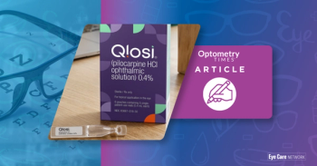
Orasis Pharmaceuticals, BlinkRx partnership offers new dispensing options for Qlosi
4
iVeena completes end-of-phase 1 FDA meeting for IVMED-85 in myopia
5