

|Articles|September 6, 2022
Corticosteroids for DED flares: safe, effective
Author(s)Cecelia Koetting, OD, FAAO, DipABO
FDA-approved steroid offers short-term relief for ocular surface inflammation.
Advertisement
Dry eye disease (DED) is a multifactorial condition. The Tear Film & Ocular Surface Society Dry Eye Workshop (TFOS DEWS) II report emphasized that the central mechanism of the disease is the loss of homeostatic tear film balance, which ultimately leads to dry eye’s key etiology, inflammation.1
The pathophysiology of DED is such that aqueous deficient and evaporative dry eye exist as a continuum; therefore, elements of each are considered in overall diagnosis and management.
In clinical practice, we see patients who have chronic dry eye as well as those who experience dry eye flares or an acute episodic worsening of symptoms owing to a variety of triggers.2-10
As our understanding has broadened regarding the nuances of the ocular surface disease (OSD) spectrum, it is becoming clear that patients with underlying chronic DED may experience acute flares and those without chronic dry eye may also suffer from flares alone.1,11,12 Inflammatory spikes of acute exacerbation occur in about 8 out of 10 dry eye patients, and about half of DED patients experience flares without continuous symptoms between 4 to 6 times per year.13-16
One of the most common triggers of acute dry eye flares in my patients is seasonal allergies, which set off inflammation and can lead to the uptick in subsequent signs and symptoms of DED. In some cases, the increased dryness stems from the systemic medications patients are taking to control their allergy symptoms.
Other triggers of acute dry eye flares include contact lens overuse, increased screen time, mask wear, autoimmune disease such as rheumatoid arthritis or psoriasis, and direct forced air from heating and cooling vents.2-10
Root of the problem
Inflammation is associated with an upregulation of intercellular adhesion molecule-1 along with matrix metalloproteinases, chemokines, tumor necrosis factor alpha, and interleukins (such as IL-1,6, and 8) in the epithelial cells of conjunctival and lacrimal tissues.17,18
These enzymes and inflammatory cells activate the inflammatory cascades inclusive of T-cell proliferation, which mediates a neurosensory response leading to the discomfort and irritation that patients experience.
The most important aspect of symptom control is getting to the root of the problem—especially in those patients with chronic disease. This means a thorough examination and workup that includes evaluating meibomian gland function, tear production, eyelid closure functionality, and the status of the lacrimal functional unit.
Ultimately, we need to proactively attack inflammation by treating the underlying condition causing the inflammation.
For our patients with both chronic dry eye and those experiencing dry eye flares regardless of chronicity, we aim for calming the inflammation before it spirals out of control. Enter topical corticosteroids, the onboarding of which is particularly useful for alleviating symptoms.
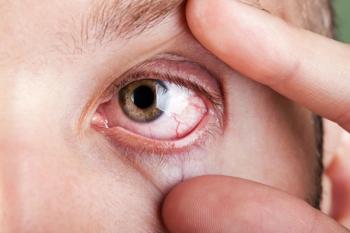
I will use a steroid in conjunction with starting a chronic medication such as cyclosporine, lifitegrast, or varenicline when indicated. Because the inflammatory cascade has already been activated, it is essential that I quell the inflammation that has flared up and is causing the increase in the patients’ signs and symptoms (Figure).
Safety, efficacy supported by large body of data
Steroids exert their effect by inhibiting upstream phospholipase A2. They block both the cyclooxygenase and lipoxygenase pathways of the inflammatory cascade, preventing formation of eicosanoids.
The agents have been shown to inhibit inflammatory cytokines, chemokines, adhesion molecules, and other inflammatory mediators.19,20 Some practitioners, however, may still be hesitant to prescribe corticosteroids for DED and other inflammatory ocular conditions because of concerns over side effects such as IOP increase and cataract formation.21,22
Corticosteroids’ safety and efficacy is well documented, specifically in reference to esters such as loteprednol etabonate, which was designed to undergo rapid metabolism.23-28
It has been shown that, in the general adult population, about one-third of people will be moderate steroid responders, experiencing IOP elevations of 6 to 15 mmHg from baseline, and 4% to 6% will be high responders, experiencing IOP elevations > 15 mmHg from baseline after 4 to 6 weeks of topical dexamethasone and/or betamethasone corticosteroid therapy.29
Steroid responders usually have predisposing factors such as a family history of glaucoma, connective tissue disease, age, diabetes mellitus, or high myopia.30,31 It is important to remember, however, that corticosteroids are backed by a plethora of research that has shown them to be effective and safe for short-term use in appropriate patients.
Today, we have an on-label topical corticosteroid specifically designed and FDA approved for the short term, up to 2 weeks, treatment of the signs and symptoms of DED—loteprednol etabonate ophthalmic suspension 0.25% (Eysuvis; ALCON).
This opens the door to give us more confidence when reaching for a steroid such as Eysuvis to manage dry eye flares. Furthermore, what separates this solution from the rest of the steroid crowd is the novel formulation utilizing Ampplify, Kala’s proprietary mucus-penetrating particle technology.
Nanoparticles of ~300 nm in diameter are coated to facilitate their penetration through the mucus barrier. This controlled delivery system enables the drop to spread more uniformly across the ocular surface.
From my clinical experience and careful review of Eysuvis data, the safety profile is extremely reassuring. Loteprednol etabonate ophthalmic suspension 0.25% was studied in more than 2,800 DED patients, and the drop was well tolerated with a low incidence of IOP increase that was similar to vehicle.
In treatment and vehicle groups respectively, 0.2% and 0% of subjects experienced a ≥ 10 mm Hg increase from baseline resulting in an IOP measurement of ≥ 21 mm Hg at any post-baseline visit up to 29 days.32-34
For practitioners who formerly had any hesitancy regarding corticosteroid use, I would say they can prescribe loteprednol etabonate ophthalmic suspension 0.25% with confidence, as it is an on-label indication supported by efficacy and safety data. I have found it to work quickly with symptom improvement starting within 48 to 72 hours, and it is a comfortable for patients to instill.
Evolution of flares conversation
Recently my discussion around acute flares of DED’s signs and symptoms has changed. We now realize that the idea of patients experiencing exacerbations makes much more sense than assuming all patients have chronic dry eye. Even my chronic dry eye patients come in at least 2 to 3 times a year, saying, “Hey, all of a sudden my eyes are worse.”
I now ask patients questions around their lifestyle to find out more about what may be triggering their dry eye flare. I let them know that having times when their symptoms get worse is normal, and I reassure them that, with treatment, the eye will calm down.
Additionally, I arm my patients by educating them that they can keep the medication on hand to use during a future flare. As part of my protocol, I see patients back to check their pressures two weeks after I start a new steroid such as Eysuvis.
Once I have IOP readings while on topical steroid in my record, I discuss with the patient that may use the topical steroid it if they have another dry eye flare twice a day for up to 2 weeks. If their symptoms do not get better or become worse, I make sure they know to call immediately. If their symptom exacerbations begin happening more often, I inform them that we need to reevaluate.
Conclusion
It is important not to minimize the side effects and risks of any medication, but rather to understand the reality and face them head on. Eye care practitioners must be diligent in their comprehensive workup of patients with OSD and identify the root cause of patients’ signs and symptoms.
Being confident in the diagnosis ensures that we are prescribing appropriate, on-target therapies that alleviate suffering. We must recognize that the majority of OSD patients have mixed mechanisms at work.
I am careful to look, lift, push, and pull the lids, examine the meibomian glands themselves, and perform meibography. Remember that both mask-associated dry eye and increased screen time are contributing to many patients’ symptoms, along with bacterial overgrowth and blepharitis.35
In most patients, the benefits of topical corticosteroids far outweigh the risks. With Eysuvis carrying an on-label FDA approved indication for the short-term treatment of the signs and symptoms of DED, there is more reason than ever to think of steroids first when patients experience present and future dry eye flares.
References
1. Craig JP, Nichols KK, Akpek EK, et al. TFOS DEWS II definition and classification report. Ocul Surf. 2017;15(3):276-283. doi:10.1016/j.jtos.2017.05.008
2. Rolando, M, Zierhut, M, Barabino, S. Should we reconsider the classification of patients with dry eye disease? Ocul Immunol Inflamm. 2021;29(3):521-523. doi:10.1080/09273948.2019.1682618
3. Amparo F, Dana R. Web-based longitudinal remote assessment of dry eye symptoms. Ocul Surf. 2018;16(2):249-253. doi:10.1016/j.jtos.2018.01.002
4. Iyer JV, Lee SY, Tong L. The dry eye disease activity log study. ScientificWorldJournal. 2012; 2012:589875. doi:10.1100/2012/589875
5. Karakus S, Agrawal D, Hindman HB, Henrich C, Ramulu PY, Akpek EK. Effects of prolonged reading on dry eye. Ophthalmology. 2018;125(10):1500-1505. doi:10.1016/j.ophtha.2018.03.039
6. Kim Y, Paik HJ, Kim MK, Choi YH, Kim DH. Short-term effects of ground-level ozone in patients with dry eye disease: a prospective clinical study. Cornea. 2019;38(12):1483-1488. doi:10.1097/ICO.0000000000002045
7. Ousler GW 3rd, Rimmer D, Smith LM, Abelson MB. Use of the controlled adverse environment (CAE) in clinical research: a review. Ophthalmol Ther. 2017;6(2):263-276. doi:10.1007/s40123-017-0110-x
8. López-Miguel, A, Tesón, M, Martín-Montañez, V, et al. Dry eye exacerbation in patients exposed to desiccating stress under controlled environmental conditions. Am J Ophthalmol. 2014;157(4):788-798. doi:10.1016/j.ajo.2014.01.001
9. Tesón M, González-Garcia MJ, López-Miguel A, et al. Influence of a controlled environment simulating an in-flight airplane cabin on dry eye disease. Invest Ophthalmol Vis Sci. 2013;54(3):2093-2099. doi:10.1167/iovs.12-11361
10. Fernández I, López-Miguel A, Enríquez-de-Salamanca A, et al. Response profiles to a controlled adverse desiccating environment based on clinical and tear molecule changes. Ocul Surf. 2019;17(3):502-515. doi:10.1016/j.jtos.2019.03.009
11. Lienert JP, Tarko L, Uchino M, Christen WG, Schaumberg DA. Long-term natural history of dry eye disease from the patient’s perspective. Ophthalmology. 2016;123(2):425-433. doi:10.1016/j.ophtha.2015.10.011
12. Perez VL, Stern ME, Pflugfelder SC. Inflammatory basis for dry eye disease flares. Exp Eye Res. 2020;201:108294. doi:10.1016/j.exer.2020.108294
13. Brazzell RK, Zickl L, Farrelly J, et al. Prevalence and characteristics of dry eye flares: a patient questionnaire survey. Presented at: AAO 2019; Oct 12-15, 2019; San Francisco, CA.
14. Brazzell RK, Zickl L, Farrelly J, et al. Prevalence and characteristics of symptomatic dry eye flares: results from patient questionnaire surveys. Poster presented at: American Optometric Association Annual Meeting 2019: October 23-27, 2019; Orlando, FL.
15. 2018 Study of Dry Eye Sufferers. Conducted by Multi-sponsor Surveys, Inc.
16. 2020 Study of Dry Eye Sufferers. Conducted by Multi-sponsor Surveys, Inc.
17. Pflugfelder SC, de Paiva CS. The pathophysiology of dry eye disease: what we know and future directions for research. Ophthalmology. 2017;124(11S):S4-S13. doi:10.1016/j.ophtha.2017.07.010
18. Gao J, Morgan G, Tieu D, et al. ICAM-1 expression predisposes ocular tissues to immune-based inflammation in dry eye patients and Sjögrens syndrome-like MRL/lpr mice. Exp Eye Res. 2004;78(4):823-835. doi:10.1016/j.exer.2003.10.024
19. Barnes PJ. Corticosteroid effects on cell signalling. Eur Respir J. 2006;27(2):413-426. doi:10.1183/09031936.06.00125404
20. Gupte R, Muse GW, Chinenov Y, Adelman K, Rogatsky I. Glucocorticoid receptor represses proinflammatory genes at distinct steps of the transcription cycle. Proc Natl Acad Sci U S A. 2013;110(36):14616-14621. doi:10.1073/pnas.1309898110
21. McGhee CN, Dean S, Danesh-Meyer H. Locally administered ocular corticosteroids: benefits and risks. Drug Saf. 2002;25(1):33-55. doi: 10.2165/00002018-200225010-00004
22. Pavesio CE, Decory HH. Treatment of ocular inflammatory conditions with loteprednol etabonate. Br J Ophthalmol. 2008;92(4):455-459. doi:10.1136/bjo.2007.132621
23. Rhen T, Cidlowski JA. Antiinflammatory action of glucocorticoids--new mechanisms for old drugs. N Engl J Med. 2005;353(16):1711-1723. doi:10.1056/NEJMra050541
24. Sendrowski DP JSD, Semes LP, Stern ME. Anti-inflammatory drugs. In: Bartlett JD, Jaanus SD, eds. Clinical Ocular Pharmacology. 5th Ed. Elsevier; 2008;12:221-244.
25. Bielory BP, Perez VL, Bielory L. Treatment of seasonal allergic conjunctivitis with ophthalmic corticosteroids: in search of the perfect ocular corticosteroids in the treatment of allergic conjunctivitis. Curr Opin Allergy Clin Immunol. 2010;10(5):469-477. doi:10.1097/ACI.0b013e32833dfa28
26. Leonardi A. The central role of conjunctival mast cells in the pathogenesis of ocular allergy. Curr Allergy Asthma Rep. 2002;2(4):325-331. doi:10.1007/s11882-002-0061-7
27. Comstock TL, Decory HH. Advances in corticosteroid therapy for ocular inflammation: loteprednol etabonate. Int J Inflam. 2012;2012:789623. doi:10.1155/2012/789623
28. Sheppard JD, Comstock TL, Cavet ME. Impact of the topical ophthalmic corticosteroid loteprednol etabonate on intraocular pressure. Adv Ther. 2016;33(4):532-552. doi:10.1007/s12325-016-0315-8
29. Armaly MF, Becker B. Intraocular pressure response to topical corticosteroids. Fed Proc. 1965;24(6):1274-1278
30. Kersey JP, Broadway DC. Corticosteroid-induced glaucoma: a review of the literature. Eye (Lond). 2006;20(4):407-416. doi:10.1038/sj.eye.6701895
31. Chang DF, Tan JJ, Tripodis Y. Risk factors for steroid response among cataract patients. J Cataract Refract Surg. 2011;37(4):675-681. doi:10.1016/j.jcrs.2010.10.051
32. Gupta PK, Venkateswaran N. The role of KPI-121 0.25% in the treatment of dry eye disease: penetrating the mucus barrier to treat periodic flares. Ther Adv Ophthalmol. 2021;13:25158414211012797. doi:10.1177/25158414211012797
33. Korenfeld M, Nichols KK, Goldberg D, et al. Safety of KPI-121 ophthalmic suspension 0.25% in patients with dry eye disease. Cornea. 2021;40(5):564-570. doi:10.1097/ICO.0000000000002452
34. Holland E, Nichols KK, Foulks GN, et al. Safety and efficacy of KPI-121 ophthalmic suspension 0.25% for DED in four RCTs. Paper presented at: American Academy of Optometry Annual Meeting 2020; November 13-15, 2020; virtual.
35. Moshirfar M, West WB Jr, Marx DP. Face mask-associated ocular irritation and dryness. Ophthalmol Ther. 2020;9(3):397-400. doi:10.1007/s40123-020-00282-6
Newsletter
Want more insights like this? Subscribe to Optometry Times and get clinical pearls and practice tips delivered straight to your inbox.
Advertisement
Related Content
Advertisement


Latest CME


Advertisement
Advertisement