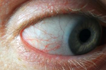
Decrease in endothelial cell density continues after iris-fixated phakic IOL explantation
The research team retrospectively studied the long-term corneal endothelial cell changes and visual outcomes after this iris-fixated phakic IOL was explanted in patients with endothelial damage and investigated any potential predictors of endothelial injury.
An iris-fixed intraocular lens (IOL) resulted in a significant decrease in endothelial cell density (ECD) 2 years after explantation,1 according to Tae Young Kim, MD, and colleagues. Kim is from the Institute of Vision Research, Department of Ophthalmology, Yonsei University College of Medicine, and the Department of Ophthalmology, Gangnam Severance Hospital, Yonsei University College of Medicine, both in Seoul, Korea.
The research team retrospectively studied the long-term corneal endothelial cell changes and visual outcomes after this iris-fixated phakic IOL was explanted in patients with endothelial damage and investigated any potential predictors of endothelial injury.
All patients had a corneal ECD below 2,000 cells/mm2 at the time of the procedure and were treated between April 2016 and October 2020. The primary study outcome was the change in corneal endothelial parameters, including ECD, over the long-term follow-up, the authors explained. Secondary outcomes included the changes in the corrected-distance visual acuity (VA) and analysis of prognostic factors.
Twenty-eight patients (44 eyes; average age, 42.5 ± 7.8 years; range, 27–63) were included in the study. The mean ECD before IOL explantation was 1,375.4 ± 468.2 cells/mm2 (range, 622–1,996), and the average duration of follow-up after explantation was 20.5 months (range, 6–58.2).
Two years after explantation, the researchers reported a more than 25% decrease in the ECD to 1,019.6 ± 368.6 mm2 (range, 608–1,689) (p< 0.01).
They also reported no significant change in the corrected-distance VA (20/23–20/22) (p = 0.59).
They found that a longer surgical duration (odds ratio, 1.004) (p= 0.04) was the only significant factor that was weakly associated with the postoperative decreases in the ECD.
They concluded that although the ECD continued to decrease despite phakic IOL explantation on a long-term follow-up, “patients did not experience any discomfort or showed decreases in VA. Therefore, a careful follow-up is required for possible endothelial injury after phakic IOL explantation.”
Reference
1. Kim TY, Moon IH, Park SE, et al. Long-term follow-up of corneal endothelial cell changes after iris-fixated phakic intraocular lens explantation. Cornea. 2023;42:150-155; doi:10.1097/ICO.0000000000003001
Newsletter
Want more insights like this? Subscribe to Optometry Times and get clinical pearls and practice tips delivered straight to your inbox.


