Defining the role of imaging in choroidal nevi
Optical coherence tomography is currently in high demand for the management of glaucoma patients, wet and dry macular degeneration, and a host of other ocular diseases.
Key Points
Choroidal nevi 101
Choroidal nevi occur in 5% to 10% of Caucasians, but are rare in dark-skinned races, according to Dr. Hessen, an instructor at Wilmer Eye Institute, Johns Hopkins University. Choroidal nevi are composed of a proliferation of spindle, polyhedral, dendritic, and balloon cells. Patients are frequently asymptomatic.
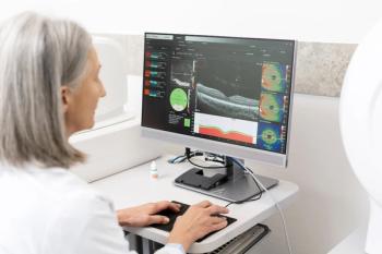
In a retrospective single-center case series of 120 consecutive eyes, researchers at Wilmer used the Zeiss Stratus OCT Model 3000 to scan 6 radial lines and complete retinal thickness analysis ( 2005;25:243-252). They analyzed the status of the retinal organization, particularly the photoreceptor layer over the nevus, and the reflectivity just posterior to the retinal pigment epithelium (RPE)/choriocapillaris and the nevus. They classified retinal edema as non-cystoid or cystoid, then compared this evaluation to a clinical evaluation in each patient.
Some findings from OCT included a more common incidence of diffuse cystoid overlying retinal edema than focal cystoid edema (8% versus 3%, respectively). For overlying retinal thickness, almost one-half were thickened (45%) compared with 32% that were of normal thickness.
"Of particular importance is that photo-receptor loss or attenuation was noted in 51% of cases. This may correspond vision loss or with visual field loss, particularly when the nevus is in or near the macula," she said.
Where OCT is more sensitive
Additional OCT findings included an increased thickness of RPE/choriocapillaris layer (68%). Optical qualities of the anterior surface were hyporeflective in 62% of nevi, and of those, 68% were pigmented and 18% were nonpigmented.
When OCT findings were compared with clinical examinations, researchers found that the OCT was more sensitive in detecting retinal edema, retinal fluid, RPE detachment, and retinal thinning.
"We were, however, better than the OCT at detecting drusen. I would say that there is a role for OCT in evaluating choroidal nevus, especially if you are not sure if there is overlying fluid present," Dr. Hessen said.
Age as a factor
The differential diagnosis for pigmented lesions includes congenital hypertrophy of the RPE, melanocytoma of the choroid, or small melanoma. For nonpigmented lesions, the differential diagnosis includes choroidal hemangioma, metastatic carcinoma, choroidal osteoma, posterior scleritis, or lymphoma.
Age may play some role in choroidal nevi formation, Dr. Hessen noted. A retrospective clinic-based study of 3,422 eyes at Wills Eye Institute reported no age-related correlation in symptoms, mean nevus base, intrinsic nevus pigmentation, related sub-retinal fluid, overlying orange pigment, RPE hyperplasia, or RPE atrophy. ( ogy. 2008;115-546-552.e2)
However, the study did find a statistical increase with age in multiple nevi per eye, mean nevus thickness, and overlying dru-sen. The investigators also concluded that the symptomatic nevi were more likely to be non-pigmented, beneath the fovea, and associated with subfoveal fluid.
Newsletter
Want more insights like this? Subscribe to Optometry Times and get clinical pearls and practice tips delivered straight to your inbox.


