Drawing the Rynerson red line: A new method to identify gland diseases
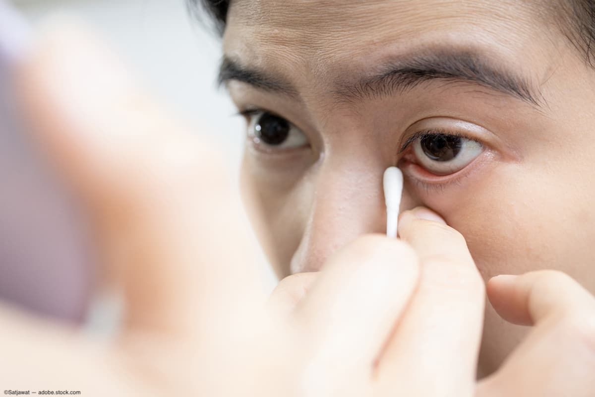
Evaluating and managing lid margin disease and meibomian gland dysfunction.


Image Credit: AdobeStock/Satjawat
Because I have 2 lid margin products commercially available, I take great interest in my professional friends and colleagues who develop tools and instruments to make the diagnosis and management of lid margin disease, dry eye disease (DED), and meibomian gland dysfunction more efficient and patient friendly. These gizmos and gadgets include but are not limited to eyelid cleaning tools and solutions, meibomian gland imagers and gland expressors, a variety of eyelid warming and hydration masks, eyelid flippers and specialty forceps, lid margin debriders, and lid/eyelash margin viewers.1
James Rynerson, MD, coauthor of the theory of dry eye blepharitis syndrome (DEBS) published in Clinical Ophthalmology in 2016, is an expert and keen observer in DED.1 The DEBS theory details how biofilm along the margin and within the structures of the eyelid precipitates chronic inflammation that slowly damages the lash follicles, meibomian glands, and (much later) the lacrimal glands, leading to what is referred to as DED or lid margin disease. A bacterial biofilm is a complex community of microorganisms, primarily bacteria, that adhere to surfaces and are embedded within a self-produced matrix of extracellular polymeric substances. The biofilm matrix consists of polysaccharides, proteins, DNA, and other molecules, creating a protective environment for the bacterial community. This protection allows bacteria to produce toxins that elicit a cycling inflammatory response.2
Rynerson is also an innovative inventor of eyelid margin disease treatment instruments that have revolutionized our daily dry eye practices. Recently, Rynerson introduced an extraordinarily simple but eloquent method to identify meibomian gland inflammation with no equipment at all, with only patient observation needed. Examining a patient for the “Rynerson red line” is an easy way to identify meibomian gland disease. This newly identified clinical entity is suggested to be a manifestation of inflammation surrounding abnormally functioning meibomian glands.
We know inflammation as an initial step in the body healing itself. Whether it is attacking an invader, such as a virus, fungus, or bacterium, or repairing a wound such as a laceration or burn, the response is generally the same. But as we know, if left unchecked, chronic inflammation can result in symptoms of discomfort, swelling, warmth, redness, and ultimately loss of function in the system it affects.3 Dilation of blood vessels allows for leakage of serum components including the important mediators of inflammation and healing (macrophages, neutrophils, growth factors, complement, dendritic cells), and an increase in redness and warmth.3
Figure 1. 1 of 2 OU photos of Rynerson red line (RRL) before multiple in-office therapy sessions (blepharoexfoliation/MG expression).
Figure 1. 2 of 2 OU photos of Rynerson red line (RRL) before multiple in-office therapy sessions (blepharoexfoliation/MG expression).
Figure 3. 1 of 2 photos of Rynerson red line (RRL) after multiple in-office therapy sessions (blepharoexfoliation/MG expression). Notice the reduced hyperemia of the RRL after treatment.
Figure 3. 2 of 2 photos of Rynerson red line after multiple in-office therapy sessions (blepharoexfoliation/MG expression). Notice the reduced hyperemia of the RRL after treatment. (Images courtesy of James Rynerson, MD)
To observe the Rynerson red line, gently stretch the excess skin on the closed eye’s upper eyelid upward and examine the area superior to the eyelash line. Rynerson maintains that a deep ruby redness traversing the eyelid that is anatomically consistent with the tracks of the meibomian glands is a reliable sign of deep and subdermal meibomian gland inflammation. The severity of this red line relates to the degree of the meibomian gland disease. The loss of meibomian gland structure appears to correlate with increase in intensity of the red line.
Figure 2. Included here is 1 of 2 OU meibography images of this representative patient. Note that posttreatment meibography in this patient did not show demonstrable change (not shown).
Figure 2. Included here is 1 of 2 OU meibography images of this representative patient. Note that posttreatment meibography in this patient did not show demonstrable change (not shown). (Images courtesy of James Rynerson, MD)
Supporting this theory, Rynerson has demonstrated improvement in the appearance of the Rynerson red line with targeted treatment of blepharitis and meibomian gland disease in his patients. The foundation of therapy for DEBS centers upon removal of biofilm from the eyelid margin and evacuation of poor quality and/or congested meibomian gland contents. Akin to anti-VEGF injections and treat-and-extend protocols for patients with retinal disease,4 the frequency and number of lid debridement and gland expression treatments until a maintenance stage is reached are individual and nuanced. Considerations that influence treatment cadence include levels of initial disease severity, strains of bacteria, pathogenicity of toxins, age, and factors that produce differing timelines for symptomatic and/or clinical improvement.


There is more to learn about the Rynerson red line. I look forward to additional exploratory and corroborative studies surrounding this clinical entity. That said, understanding the mechanisms of biofilm formation and strategies to disrupt or prevent biofilm formation is essential for developing effective treatments to prevent the downstream deleterious effects of bacteria-based disease.
References:
Zhang L, Wang J, Gao Y. Eyelid cleaning: methods, tools, and clinical applications. Indian J Ophthalmol. 2023;71(12):3607-3614. doi:10.4103/IJO.IJO_1457_23
Rynerson JM, Perry HD. DEBS - a unification theory for dry eye and blepharitis. Clin Ophthalmol. 2016;10:2455-2467. doi:10.2147/OPTH.S114674
What is an inflammation? In: Institute for Quality and Efficiency in Health Care. InformedHealth.org. Institute for Quality and Efficiency in Health Care; 2006. Updated February 22, 2018. Accessed March 8, 2024. https://www.ncbi.nlm.nih.gov/books/NBK279298/
Skelly A, Bezlyak V, Liew G, Kap E, Sagkriotis A. Treat and extend treatment interval patterns with anti-VEGF therapy in nAMD patients. Vision (Basel). 2019;3(3):41. doi:10.3390/vision3030041