'First treatment for keratoconus itself
About two in 10 patients with keratoconus requires eventual corneal transplantation, and these patients comprise 5,000, or 15%, of the corneal transplants in the United States each year.
New York-About two in 10 patients with keratoconus requires eventual corneal transplantation, and these patients comprise 5,000, or 15%, of the corneal transplants in the United States each year, according to William Tullo, OD, FAAO, senior director of optometric services at TLC Laser Eye Centers. Even so, corneal transplants aim to restore vision, not treat the underlying disease that necessitated the transplant.
"One of the most exciting procedures soon to be available to optometrists and ophthalmologists in the United States" is how Dr. Tullo described CXL at International Vision Expo East here in March.
Given that some 70% of patients with keratoconus first present to optometric practices, ODs can play an important role in referring patients to approved trial sites.
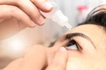
Simply put, CXL, which first had applications in dentistry, combines riboflavin drops (vitamin B2) and 370nm ultraviolet A light (UVA). The resulting interaction forms chemical bonds that stiffen the collagen fibrils, making the cornea biomechanically stronger.
"What is happening in the cornea is actually covalent bonding," Dr. Tullo said. "Instead of the typical mindset of using anti-oxidants to reduce irritation to the body, we're actually using oxidants," he said. "Riboflavin acts as a photosensitizer producing free-oxygen radicals that are produced by the combination with ultraviolet A light, causing covalent bonding between adjacent collagen fibrils via the lysyl oxidase pathway."
Besides keratoconus, CXL is indicated for treating other ectatic conditions of the cornea, including pellucid marginal degeneration, keratoglobus, and forme fruste keratoconus. It also can be used to stabilize corneas with surgically induced weakening, including that caused by post-LASIK and post-PRK ectasia or post-RK induced variable vision.
One thing CXL cannot currently accomplish is to treat refractive error or predictably provide good uncorrected visual acuity. However, CXL can normalize the corneal shape and reduce irregular astigmatism so that the patient can wear glasses and be fit with contact lenses more easily. "The goal of this is to stop the progression of keratoconus; to make it a lot easier to refract the patient at the phoropter," he said.
The Dresden protocol
One procedure moving through FDA trials uses the Dresden protocol, the first protocol developed to do CXL (though not the one primarily used outside the United States).
The Dresden protocol begins with pachymetry, which is essential to ensure corneal thickness of more than 400µ. If the cornea is less than 400µ thick, the patient is at risk for developing corneal haze. This emphasizes the importance of referring patients early in the disease process, possibly as young as 10 or 11, before their corneas thin and vision significantly declines.
The surgeon then removes the central corneal epithelium then instills 1 drop of riboflavin 0.1% every 2 minutes for 30 minutes. The riboflavin, which comes in a dextran-based solution, protects against excessive UV exposure to the posterior structures of the eye.
After 30 minutes and the slit lamp examination ensures that the riboflavin has significantly and uniformly penetrated the stroma, the surgeon aims the photomediator at the central 6mm of the cornea. Total exposure is 30 minutes, or about 3 mW/cm2. During the UV exposure a technician adds 1 drop of riboflavin every 2 minutes.
At the end of the procedure, the surgeon places a bandage contact lens on the patient's eye for 4 to 5 days while the cornea re-epithelializes. Perioperative care is similar to PRK patients requiring a regimen of a topical fluoroquinolone and steroid.
Newsletter
Want more insights like this? Subscribe to Optometry Times and get clinical pearls and practice tips delivered straight to your inbox.


.png)


