

|Articles|July 20, 2023
- July digital edition 2023
- Volume 15
- Issue 07
Fitting scleral lenses for Bell palsy and Ramsay Hunt syndrome
Author(s)Jon Bundy, OD
Well-fitting lenses greatly improve quality of life for patients.
Advertisement
Bell palsy, and its less common counterpart Ramsay Hunt syndrome, are well known to primary eye care providers. Both maladies cause significant negative impacts on quality of life, especially when the ocular surface is involved. The following review presents 3 separate but related cases in which each patient’s quality of life was greatly improved with the addition of a scleral lens.
LL, a 72-year-old white woman, presented to our office with the right side of her face drooping since she woke up that morning. She had undergone radial keratotomy surgery in both eyes 30 years prior and was taking brinzolamide 1% ophthalmic suspension (Azopt) twice daily OD for mild glaucoma. Two days prior, her primary care provider prescribed oral cephalexin for an ear infection. She was given a tentative diagnosis of Ramsay Hunt syndrome and offered palliative measures, including a bandage soft contact lens (Biofinity) and recommended preservative-free lubricating drops OD every hour.
She returned a week later having visited the local emergency department, where they confirmed she did not have a stroke and prescribed a 10-day course of oral prednisone. After another week, LL was struggling with vertigo, had “cried out” the contact lens the day prior, and was wearing a patch to keep her right eye closed. The right cornea now demonstrated numerous filaments. Patients with Ramsay Hunt syndrome often have more severe sequelae and are less likely to recover than patients with Bell palsy.1 Given the escalation of signs and symptoms, a scleral shell was recommended for more aggressive treatment and a more long-term plan.
The Ampleye from Art Optical was selected with the 4000-µm sagittal height. Central clearance was 307 µm after 20 minutes of settling. Peripheral edges were fitting properly as they sat 50% into the surrounding conjunctiva. There was no NaFl uptake, but there was significant conjunctival prolapse under the scleral lens 360 degrees around. The limbal vault was deemed too high, causing excess suction, so it was reduced. The lens was ordered in Optimum Extra material with Tangible Hydra-PEG coating to maximize lubrication and comfort. At dispense, the lens was fitting perfectly, with no conjunctival prolapse. Three years later, LL is still discouraged that her face remains slightly disfigured from the paralysis, but her ocular surface remains stable and she has maintained good vision by using the scleral lens.
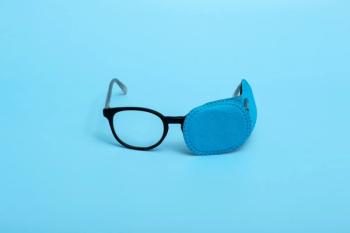
JL, a 66-year-old white woman, presented to our office with a dry, painful left eye for the past 7 years after an episode of Bell palsy (Figure 1). JL showed moderate lagophthalmos of both the upper and lower left eyelids, with significant corneal staining and conjunctival injection. She was tired of the constant pain, fluctuating vision, and recurring infections and was hoping for a better solution than administering lubricating drops every day and ointment at night.
The Custom Stable Elite from Valley Contax was selected, and a 4310-µm sagittal height diagnostic was selected. The manufacturer recommends converting flat keratometry (K), or the flattest cornea meridian—as measured by topography or auto-keratometry in diopters (D)—to microns for the initial lens, and JL’s keratometry measurements were 43.75/44.75 D. The lens showed central corneal touch, so a steeper lens was selected. The 4570-µm lens showed good central clearance of 165 µm after 20 minutes of settling. Peripheral edges were 1 step loose in the steep meridian, allowing NaFl under the lens; this could eventually cause midday fogging, which is a common complaint of scleral lens wearers.2 To combat the peripheral edge lift, the toricity of the lens was increased 1 step. The lens was ordered in Optimum Extra material with Tangible Hydra-PEG coating (Figure 2).
Six years after our initial visit, JL continues to wear her scleral lens every day. We replace it every 2 years or so as it gets scratched and starts to accumulate deposits.
CL, a 75-year-old white woman, presented to our office with a lifetime history of ocular complications. She was diagnosed with Bell palsy on the right side of her face at approximately 3 months of age. It is one of the most common neurological disorders in children, and the diagnosis is confirmed after all other forms of acute peripheral palsy have been excluded.3 She had facial sling surgery at age 26, as well as a face lift, brow lift, and blepharoplasty at age 50. At age 68, a gold weight was placed in her right upper eyelid to try to facilitate a more normal blink response.
CL was battling chronic infections in her right eye as well as chronic inflammation at the time of presenting to our office. Without using prednisolone acetate ophthalmic suspension, 1% every other day OD, that eye would become extremely painful with unusable vision. With the steroid, her IOP would rise above normal but fortunately had not caused any optic nerve damage yet. CL would patch the eye at night, which caused recurrent infections for which she would use tobramycin drops “a few times per day for a few days,” in addition to the preservative-free lubricating drops she was using multiple times every day. She had near constant trichiasis of the lower eyelid. We would generally not epilate the lashes as they were often long enough to lay gently along the cornea, whereas epilation would bring relief for a week or 2 and then the lashes would be very short and sharp as they regrow.
The Custom Stable Elite from Valley Contax and a 4570-µm sagittal height diagnostic were selected. The manufacturer recommends converting flat K to microns for the initial lens, and CL’s keratometry measurements were 44.00/47.25 D. Central clearance was higher than ideal at 635 µm, and the peripheral edges fit very well with no edge lift and no conjunctival impingement. A 4310-µm diagnostic lens was then selected, which gave better central clearance at 428 µm but still higher than preferred. The peripheral edges were 1 step flat and allowed NaFl under the lens. Adjustments were made to lower the central clearance (limbal clearance zone +1) and tighten the peripheral edges (scleral landing zone –1). The lens was made to achieve a +1.00 DS over-refraction so CL could continue to wear her eyeglasses prescription with a –1.00 DS lens OD.
CL quickly adapted to the new lens and was able to discontinue the prednisolone drops. Over the past 7 years of wearing the lens, she has averaged less than 1 occurrence of conjunctivitis per year. After wearing a scleral lens OD for 5 years, CL presented to our office, explaining that she thought her “gold weight was coming out of her eyelid.” Sure enough, the gold weight that was implanted 14 years prior was now visible anteriorly through her upper eyelid (Figure 3). We scheduled an evaluation with our preferred oculoplastic specialist.
CL returned 3 weeks later very concerned because she could now see the gold weight protruding (Figure 4). We expedited the oculoplastic consultation, and she was seen within the week.
The gold weight was removed without complication and CL healed with dissolvable sutures closing the open wound (Figure 5). It is reasonable to assume the scleral lens exacerbated the gold weight extrusion, as the lens could conceivably create a posterior to anterior force onto the upper eyelid as compared with a relatively soft and flexible cornea. For this reason, I will discuss this possibility with any future patient but will not treat it as an absolute contraindication. CL had no concern with the complication as, in her words, “the scleral lens has given me my life back.”
Inserting a scleral lens is often the greatest barrier to entry for wearing the lens. Fortunately, in patients with Bell palsy, the lagophthalmos of the upper and lower eyelids creates a larger than normal aperture, so inserting the lens is often easier in these patients. This is especially helpful in older patients who may have lost some dexterity over the years. Please don’t discount the idea of a scleral lens in these patients simply due to advancing age. All 3 patients presented here have not only shown dramatic dry eye disease improvement, but they have also shown significant quality of life improvement with their scleral lenses.
References
1. Sweeney CJ, Gilden DH. Ramsay Hunt syndrome. J Neurol Neurosurg Psychiatry. 2001;71(2):149-154. doi:10.1136/jnnp.71.2.149
2. Fogt JS. Midday fogging of scleral contact lenses: current perspectives. Clin Optom (Auckl). 2021;13:209-219. doi:10.2147/OPTO.S284634
3. Khair AM, Ibrahim K. Idiopathic non-traumatic facial nerve palsy (Bell’s palsy) in neonates; an atypical age and management dilemma. Oman Med J. 2018;33(1):65-68. doi:10.5001/omj.2018.12
Articles in this issue
almost 3 years ago
What is type 3 diabetes?almost 3 years ago
Demodex “tails”almost 3 years ago
Pediatric myopia and keratoconus: Key detection and treatment strategiesalmost 3 years ago
Dry eye after LASIK is a common problemAdvertisement
Related Content
Advertisement


Latest CME


Advertisement
Advertisement
Trending on Optometry Times - Clinical News & Expert Optometrist Insights
1
Study finds no link between semaglutide and a blinding form of macular degeneration
2
Euclid Vision receives FDA clearance for Be Free Day toric contact lenses
3
Oculis enrolls first patient in licaminlimab PREDICT-1 DED trial
4
Beyond visual acuity: BostonSight's current research into mental health and lid disease
5