High IOP, corneal edema with unknown etiology
A few days ago, a 61-year-old white male presented with a gradual and painless loss of vision in his left eye.
Since the dawn of humanity, the question concerning from whence we came has been one of the most pondered ideals of our species. Almost every so-called “great thinker,” from the pre-Socratics to Sartre, has had something to say on the matter.
Throughout civilized history, philosophers, theologians, and the like have attempted to nail down this elusive notion. So, how does this tie in with glaucoma? Let’s see.
Case presentation
A few days ago, a 61-year-old white male presented with a gradual and painless loss of vision in his left eye. His medical history was remarkable for arterial hypertension, high cholesterol, type 2 herpes simplex virus, and “pre-glaucoma.”
His medications included hydrochlorothiazide (Esidrix, USP), simvastatin (Zocor, Merck), valacyclovir (Valtrex, GSK), and latanoprost (Xalatan, Pfizer), which he was taking every morning. He stated that the vision in his left eye got a little foggy four days prior and had gotten progressively worse since then with no redness or pain. He had not attempted any treatment before presenting to me.
Related:
Entering acuities through his habitual spectacle correction were 20/25 in the right eye and 20/400 in the left eye with no improvement upon pinhole testing. Lensometry showed his current spectacle correction to be -0.50 DS in the right eye and -0.75-0.50 x 117 in the left eye with a +2.00 D add in each eye.
Pupil function was unremarkable in each eye with neither pupil reacting sluggishly. Extraocular muscle testing was normal in each eye with no pain, as was confrontation visual field testing.
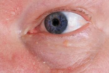
Slit lamp examination showed grade-4 microcystic edema of the left corneal epithelium with Grade 2 folds in Descemet’s membrane. The edema was not confined to the central portion of the cornea but was rather diffuse all the way out to the limbus in all directions.
The anterior chamber of the left eye was difficult to view, but there were no frank signs of active or past inflammation. The anterior segment of the right eye was unremarkable, and both angles were open by the von Herrick method.
Gonioscopy of the right eye showed all four quadrants open to the ciliary body with a flat iris approach and Grade 1 pigment in the trabecular meshwork. Gonioscopy of the left eye was obscured by the corneal edema with angle structures posterior to Schwab’s line grossly visible.
Intraocular pressures (IOPs) by Goldmann applanation tonometry were 24 mm Hg in the right eye and 53 mm Hg in the left eye at 10:15 a.m. Undilated assessment of the right optic nerve showed a moderate sized disc with an intact neuroretinal rim and no frank defects. The left posterior segment was obscured.
Treatment
I explained to the patient that his loss of vision was due to swelling of his cornea from very high eye pressure and that we needed to lower his eye pressure in order to get him seeing better and also to assess what else may be going on with that eye.
I instilled two drops of Combigan (brimonidine tartrate/timolol maleate, Allergan) into the left eye and instructed the patient to return in two hours for follow-up care. When he returned, the vision in his left eye had improved to 20/40, and his IOP in that eye had improved to 26 mm Hg.
Remarkably, the corneal edema in the left eye had almost resolved completely. The left anterior chamber was deep and quiet, and dilated fundoscopy showed unremarkable posterior segments and healthy optic nerves with cup-to-disc ratios of 0.2 x 0.2 vertically and horizontally in each eye.
I instructed the patient to discontinue his latanoprost and sent him home with a sample of Combigan for use every morning and afternoon in both eyes.
Related:
Four days later, his IOPs were 16 mm Hg in the right eye and 18 mm Hg in the left eye at 11 a.m. with complete resolution of the corneal edema. At that point, the patient informed me that since he worked just a few blocks down from me, he wanted to come to me for his continued care.
I invited him back in two weeks for baseline glaucoma testing and am currently waiting on his medical records so that I can see from whence he came. I am particularly interested in seeing his highest IOPs and why he is using latanoprost in the morning. However, I am more interested in seeing if anything like this has happened to him before.
Herpes and ocular hypertension
It is intriguing that the patient has a history of herpetic disease (although he could remember no herpetic eye issues). The herpes simplex virus, in its various types, can cause ocular hypertension through exacerbated inflammation of the anterior chamber and trabecular meshwork.1
The resultant ocular hypertension can cause corneal edema, but I would have expected to see at least a mild anterior chamber reaction if the herpes simplex virus was the cause of all of this. At this point, I am treating this as an isolated occurrence and am labeling this patient as having ocular hypertension.
All he could tell me about his ocular history was that he was diagnosed with “pre-glaucoma” and told to take drops for it. I will watch him closely over the next several weeks, but I am eager to get his records in hopes of better deciphering how his IOPs and anterior segments have behaved in the past.
Related:
References:
1. Nalcacioglu-Yüksekkaya P, Ozdal PC, Teke MY, et al. Presumed herpetic anterior uveitis: a study with retrospective analysis of 79 cases. Eur J Ophthalmol. 2014 Jan-Feb;24(1):14-20.
Newsletter
Want more insights like this? Subscribe to Optometry Times and get clinical pearls and practice tips delivered straight to your inbox.


