
|Articles|December 16, 2014
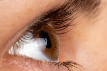
The importance of pachymetry and CCT
Reasons for thin CCT values as independent predictors of POAG have been relatively unclear, and there aren’t nearly as many studies concerning CCT as there are concerning risk factors such as IOP.
Advertisement
The measurement of central corneal thickness (CCT) by pachymetry has been an essential part of the contemporary
Reasons for thin CCT values as independent predictors of POAG have been relatively unclear, and there aren’t nearly as many studies concerning CCT as there are concerning risk factors such as IOP. Moreover, the definitive answer to the question of a causal vs. correlative relationship of CCT to POAG remains somewhat elusive. However, one study recently published in the American Journal of Ophthalmology may help to shed some light on this intriguing feature of glaucoma.3
Oxygen levels of the anterior chamber
In this cross-sectional study, 124 patients who were about to undergo cataract or glaucoma surgery had the oxygen levels of their anterior chambers measured directly by means of paracentesis and a fiber optic oxygen sensor at three different and distinct locations: just posterior to the central corneal endothelium, midway through the anterior chamber, and at the anterior chamber angle. The results of the study showed that oxygen levels were higher in patients with thinner CCT values. However, this result was isolated to the anterior chamber angles and not the other sites in the anterior chambers measured in these patients.
Such a specific correlation is particularly intriguing due to the fact that the trabecular meshwork is located within the anterior chamber angle. Could it be that oxygen and/or reactive oxygen species may damage the trabecular meshwork on a molecular level, thus affecting its structural, and therefore, functional integrity? The authors of the study are among those who suggest this very possibility, and their results certainly provide some evidence for such a scenario.
Oxidative stress from oxygen and/or reactive oxygen species resulting in tissue damage is not a novel concept in the arena of ocular disease (or glaucoma, specifically, for that matter). From the cornea to the choroid, just about every tissue in the human eye is susceptible to damage from a hyperoxic state. The Age-related Eye Disease Study (AREDS) has clearly demonstrated the effectiveness of antioxidant supplementation in patients with macular degeneration.4 Oxidative stress likely often plays a role in glaucoma development and progression as well, and, with this in mind, certain novel therapeutic molecules, such as Rho kinase (ROCK) inhibitors, are being investigated as potential neuroprotective agents in the fight against glaucoma.5 It has been shown that oxidative stress plays a significant role in damage to the trabecular meshwork in glaucoma models.6 Since the trabecular meshwork and the lamina cribosa share common derivation from the neuro-ectoderm, there may be good reason to believe that oxidative stress would damage the lamina cribosa in a similar fashion, thus exposing and predisposing the retinal ganglion cells that fenestrate through it to damage and eventual premature cell death (as happens in glaucoma).
The importance of pachymetry
So, with the potential relationship between CCT values and oxidative stress (and the OHTS study) in mind, pachymetry, while relatively simple and straightforward to perform, isn’t merely the mundane and boring task we think it is. In fact, reliable pachymetry values are so important and essential to an accurate glaucoma work-up that it’s one of the few ancillary tests that I insist on performing myself (rather than delegating to a technician). It’s just too important, as I can recall a couple of patient scenarios in which thin CCT values were the tipping point for treating rather than monitoring for change.
Further, the suggested inverse relationship between CCT values and oxygen levels in the anterior chamber angle acts as another check in the box that we, as clinicians, are truly on the verge of being able to treat glaucoma in ways other than lowering IOP. The potential for these concepts to lead to such new horizons in glaucoma treatment is especially exciting in the presence of conditions such as normal-tension glaucoma, in which IOP plays a role, but perhaps less of a role. Thinking about glaucoma variables other than IOP as risk factors also helps us, as clinicians, to move beyond that golden number of 21 mm Hg.
The future of
So, in the age of big and colorful OCT printouts that quantify retinal nerve fiber and ganglion cell layer thickness down to just a few microns, I think it’s pretty cool that little ol’ pachymetry is still so important.
References
1. Care of the Patient with Open Angle Glaucoma. The American Optometric Association Clinical Practice Guideline (revised 2010). Available at:
2. Brandt JD, Beiser JA, Kass MA, et al. Central corneal thickness in the Ocular Hypertension Treatment Study (OHTS). Ophthalmology. 2001 Oct;108(10):1779-88.
3. Siegfried CJ, Shui YB, Bai F, et al. Central Corneal Thickness Correlates with Oxygen Levels in the Human Anterior Chamber Angle. Am J Ophthalmol. 2014 Nov 26.
4. Chew EY, Clemons TE, Agron E, et al. Long-term effects of vitamins C and E, β-carotene, and zinc on age-related macular degeneration: AREDS report no. 35. Ophthalmology. 2013 Aug;120(8):1604-11.
5. Yamamoto K, Maruyama K, Himori N, et al. The Novel Rho Kinase (ROCK) Inhibitor K-115: A New Candidate Drug for Neuroprotective Treatment in Glaucoma. Invest Ophthalmol Vis Sci. 2014 Oct 2;55(11):7126-36.
6. Sacca SC, Pulliero A, Izzotti A. The dysfunction of the trabecular meshwork during glaucoma course. J Cell Physiol. 2015 Mar;230(3):510-25.
Newsletter
Want more insights like this? Subscribe to Optometry Times and get clinical pearls and practice tips delivered straight to your inbox.
Advertisement
Related Content
Advertisement


Latest CME


Advertisement
Advertisement
Trending on Optometry Times - Clinical News & Expert Optometrist Insights
1
The top stories in glaucoma to enter the new year with
2
Euclid Vision Corporation launches new Be Free Day lens for myopia management
3
GSLS 2026: Enhancing visual quality in keratoconus
4
GSLS 2026: Keratoconus lens fitting before and after surgery
5