

|Articles|November 20, 2015
An OD’s perspective on his own cataract surgery
For years, I have been an advocate of early cataract surgery in any symptomatic patients. As we all know, the progression of cataract development is an unavoidable process, so why delay the inevitable?
Advertisement
For years, I have been an advocate of early cataract surgery in any symptomatic patients. As we all know, the progression of cataract development is an unavoidable process, so why delay the inevitable? You get the ability to plan future refractive error, to minimize visual symptoms and lifestyle restrictions, and to pre-plan a convenient time and schedule.
Related:
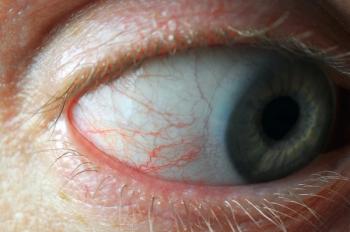
Last fall, at age 69, I was in the position of being able to follow my own best advice. Although my best-corrected vision under optimal conditions was still in the 20/25 range, my contrast sensitivity was noticeably reduced, and my best-corrected acuity was reduced to 20/40 under glare situations. I was also experiencing an increase in streaks and halos around lights at night, as well as a need for more and more illumination for visual chores like color coordination and fine detail close work.
Katherine Mastrota, OD, FAAO, center director at Omni Eye Surgery, worked with me through my cataract surgery process from initial evaluation through post-operative care. My surgeon was Douglas Grayson, MD, Omni’s cataract specialist. Dr. Mastrota and I thought it would be interesting for me to share my personal and professional observations on a process that until now I have seen only from the other side of the slit lamp biomicroscope.
Related:
Decisions for the cataract patient
As we all know, there are a number of decisions that must be made before cataract surgery can take place. Before comanagement, the surgeon typically chose the refractive outcome for the patient, usually deciding on bilateral distance vision. In the modern practice dynamic, this decision is now shared between doctor and patient. In my case, they were one and the same. As a lifelong myope, I have always understood the benefits of myopia, even more so as a presbyope. I wear contact lenses for sports, athletics, and some social activities, but single-vision distance correction has been a frustrating experience since my mid 40s. I reasoned that the frustration of poor acuity at near would be even more troublesome with no accommodation. Having personal experience with monovision, I chose it as my refractive goal, understanding that there is no perfect substitute for the young, clear, healthy, accommodating human eye.
The next decision to be made is tried and true vs. new and sexy; or more technically put, phacoemulsification vs. femtosecond laser-assisted cataract surgery. Medicare and other insurers have so far considered femto to be an upgraded, non-essential procedure, and has made it an out of pocket option for patients. Many ophthalmological surgery centers have chosen to incorporate the laser fee into the price of upgraded toric or multifocal intraocular lenses (IOLs), removing the choice of femto vs. phaco from the equation. In my case, needing only spherical monofocal IOLs, I could choose either procedure.
Related:
I am frequently asked to help my patients choose the right procedure for them. Frankly, in many cases, the real concern is whether the new laser treatment is worth the difference in cost. Every ophthalmologist performs phaco, and most have probably never done anything else. If you, as a professional, feel that femto is in your patient’s best interest, and there are many reasons why it might be, you should consider steering your patient to a surgeon who can offer either procedure, like Dr. Grayson.
I realized that I was in a unique position to compare the two procedures from a patient’s perspective, so I opted to have one of each.
Phaco in the left eye
My left (dominant) eye was operated on first, using phacoemulsification, aimed at plano.
The procedure itself was smooth and without pain, thanks to the IV sedatives. The post-op healing process, on the other hand, was a little worrisome.
Initially, things seemed hazy and watery, as if the media had been replaced with something cloudy. At my one-day post op I had a steamy cornea and an intraocular pressure (IOP) of 41 mm Hg, which was not alarming to Dr. Mastrota, and which would not have been alarming to me had it been in someone else’s eye, but this was in mine! My IOP dropped to the low 20s in one hour after a pressure-lowering drop, but I’d guess that to most patients that would be a very long hour. Virtually all the haze disappeared when the IOP normalized, but the media itself took four days to truly clear. This was due to the visco-elastic material injected into the anterior chamber during IOL implantation working its way out through the trabecular meshwork. Most patients would think of this as a disturbing complication and would breathe a sigh of relief when it dissipated, as did I. At the end of four days, my uncorrected VA was 20/25.
More from Dr. Klein:
Femto in the right eye
My right eye was operated on two weeks later using femtosecond laser assistance, aimed at -1.75 D.
The vision in the femto eye cleared in one day, with few, if any, disturbances. IOP never went above 20 mm Hg, and there was no noticeable change in media clarity.
In the interest of transparency (no double meaning intended), I do have pseudoexfoliation syndrome (PXF), and while Dr. Mastrota thought there was more pre-op pigment in the angle of the left eye (and we know that PXF eyes generally have a robust inflammatory response to surgery), I can’t believe that the difference in visco-elastic clearing was solely attributable to the difference in PXF. Thinking as any patient would, I reasoned that the difference in the procedure itself must have been a factor.
Related:
Comparing procedures
As a consequence of scheduling, each surgery was done in a different facility. The first surgery was done with a nerve block. The immediate results of the nerve block were absolutely frightening. Because the nerve block also affects the extraocular muscles and the orbicularis, I noticed, for hours after the surgery, very significant double vision. The diplopia was both horizontal and vertical, and I couldn’t close the operated eye to alleviate it. In addition, the image in the operated eye was tilted about 30 degrees. I think I remember being taught in optometry school that the human eye does not cyclorotate, but let me tell you, it sure seemed like it did.
Related:
None of these phenomena occurred with the second eye, which was anesthetized with topical anesthetics only. In both eyes, there was no noticeable post-op pain and no significant difference in dry eye perception. If given a choice, I’d pick topical drops over nerve block any day.
Femto also offers lowered retinal trauma risk, better capsulorhexis centration, and the ability to incorporate astigmatic keratectomy incisions. In the hands of a surgeon skilled with the laser, I’ve yet to hear a clear argument against it. If cost and access are not an issue, my advice to patients, now based on personal experience, will be to choose laser-assistance.
As an addendum, I should mention that because I opted for early intervention, there was no “a-ha” moment in which colors looked substantially more vivid, and background light looked substantially brighter. A patient with BCVA of 20/200 will have much more of a “wow” factor.
Related:
Most people with aging eyes, even eyecare professionals, have a difficult time separating the visual disturbances caused by cataracts from those caused by vitreous opacities and irregularities. My floaters are even more apparent now. I plan to be a lot more diligent, going forward, in stressing to patients that they are getting a new crystalline lens, not a new eyeball.
So here I am, six months out from my cataract surgeries, functioning nicely with my new vision and pleased to know that this is one aging issue that I have already put to rest. If the others fix up this handily, I’ll be one happy old camper.
Newsletter
Want more insights like this? Subscribe to Optometry Times and get clinical pearls and practice tips delivered straight to your inbox.
Advertisement
Related Content
Advertisement


Latest CME


Advertisement
Advertisement
Trending on Optometry Times - Clinical News & Expert Optometrist Insights
1
Rapid ocular surface optimization in pre-op patients with cataract
2
Deseyne EDOF daily contact lenses first of its kind approved by US FDA
3
What’s new in 2025-2026: Ophthalmic drugs and drug delivery systems
4
PRIMA retinal implant restores vision in patients with advanced GA
5