|Articles|July 20, 2017
Reviewing anterior segment ARVO 2017 posters
So much basic science research is presented at this meeting, and most of it will be years before it makes its way to clinical trials. Let’s concentrate on research that might be of use to us in the exam room very soon.
Advertisement
Like most of us, I go to my fair share of optometry meetings each year. I've come to know how optometric meetings flow. The Association for Research in Vision and Ophthalmology (ARVO) format is different from anything you'll see at a conventional optometric meeting.
The massive room includes hundreds upon hundreds of posters with groups of people gathered around the posters in intense study. Critical observation was the norm. I've never seen as many notebooks out and notes feverishly being taken or researchers of like interests sharing and discussing each other's work openly and freely.
Previously from Dr. Bowling:
With so much information being presented in so many different research areas, there is no way any one person could cover all the topics. Fortunately, Editorial Advisory Board member Stuart Richer, OD, FAAO covered the retina and posterior segment in an earlier feature. (
I will focus my attention on anterior segment presentations, particularly on clinical research. So much basic science research is presented at this meeting, and most of it will be years before it makes its way to clinical trials. Let’s concentrate on research that might be of use to us in the exam room very soon.
Cornea
One of the first posters I observed discussed a new measurement of tear film break-up time based on corneal reflex interference patterns. This work by Mikel Aldaba, BSc Optom, PhD, and associates from Spain demonstrated in 10 subjects a means of measuring tear break-up time non-invasively using interference patterns. Results demonstrated the break-up time measurement of the tear film was similar to that found using fluorescein. #471
Another poster I found very timely from second-year medical student Owen Drinkwater and colleagues from the Cornell School of Medicine assessed the prevalence of abnormal tear testing in cataract surgery patients using both TearLab Osmolarity System and InflammaDry (Quidel). They found that 43 percent of test subjects had both a positive tear osmolarity test and a positive MMP-9 test prior to cataract surgery. As ocular surface disease (OSD) of any severity level can lead to adverse outcomes in cataract and refractive surgery, point-of-care diagnostic testing may be useful in identifying early or asymptomatic OSD and pre-operative cataract surgery patients, improving refractive outcomes. #791
Related:
Perhaps because one of my patients recently presented with a particularly recalcitrant recurrent erosion (RCE), I found a poster by Optometry Times Editorial Advisory Board members Scott Hauswirth, OD, FAAO, and Milton Hom, OD, FAAO to be helpful. Performing a retrospective review of 53 RCE cases, the authors developed an algorithm for treating RCEs.
On first occurrence or presentation in which the RCE component is questionable, use lubrication, hyperosmotics, and a bandage contact lens. On a second occurrence with a high suspicion of RCE, discuss and consider the use of epithelial debridement along with a cryopreserved amniotic membrane. On third occurrence in which RCE diagnosis is confirmed, perform epithelial debridement and use a cryopreserved amniotic membrane. The use of amniotic membranes is growing in optometry, especially in conditions such as these, so it is important to define when such measures should be used. #2612
In updated surveillance data from the Antibiotic Resistance Monitoring in Ocular Microorganisns (ARMOR) study, 359 isolates were collected that were susceptible to all antibiotics tested. While resistance to Pseudomonas aeruginosa isolates continues to be low, non-susceptibility to fluoroquinolones (7 percent) more than doubled from 2015. Isolates of Streptococcus pneumoniae exhibited non-susceptibility to azithromycin (31 percent) and penicillin (38 percent) while remaining susceptible to fluoroquinolones. Among all staphylococci, resistance was most notable for azithromycin (47 to 63 percent), oxacillin/methicillin (27 to 43 percent), and ciprofloxacin (25 to 30 percent). Bacterial resistance among micro-organisms continues to grow, and clinicians must follow accepted practices to limit the increase of antibiotic resistance. #1083
Related:
After a long period without new topical ophthalmic antibiotics, new drugs are on the horizon.
Pazufloxacin 0.09% (Laboratories Sophia) in different dose regimens showed similar bacteriological and clinical efficacy when compared to moxifloxacin (Vigamox, Alcon) and gatifloxacin (Zymar, Allergan) in patients with bacterial conjunctivitis in a multicenter double-blind clinical trial of 300 patients. #1088
Another presentation showed solithromycin (Cempra), a next-generation macrolide, to be 8 to 16 times more potent than azithromycin in vitro and is active against azithromycin-resistant strains. Solithromycin has the potential to treat a variety of ocular infections, as well as ocular surface disease such as meibomian gland dysfunction and blepharitis. #1089
Any of us who have dealt with recurrent uveitis know how frustrating it can be for both the clinician and the patient. One poster showed the amazing effect of 50,00 IUs of vitamin D administered weekly. In 35 patients with relapses of their uveitis treated with this dosage of vitamin D, none showed a relapse in their uveitis in a year of therapy. #160
After vitamin D treatment, there was also an improvement in symptoms and signs normally associated with uveitis relapses, including ocular pain, blurring of vision, pericorneal hyperemia, and aqueous or vitreous cells or flare. Vitamin D has been shown as an essential organic compound which has a crucial effect on the immune response. Another study showed that a lack of Vitamin D contributes to corneal neuropathy in Sjögren syndrome patients. #1018
Related:
Dry eye
Dry eye research continues to grow in the ophthalmic community as evidenced by the number of posters addressing the topic.
Perhaps one of the most telling posters discussed dry eye practice patterns. The study from Scheie Eye Institute and Wills Eye Hospital surveyed 101 ophthalmologists, which included 43 corneal specialists, for their practice patterns regarding the evaluation of dry eye patients. The top three most common traditional dry eyes tests performed were corneal fluorescein staining (89 percent), tear break-up time (78 percent), and anesthetized Schirmer’s test (51 percent).
Conjunctival lissamine green and/or rose bengal staining were performed by less than 25 percent of respondents. The top three newer dry eye tests performed were tear osmolarity assessment (23 percent), MMP-9 testing (17 percent) and LipiView (TearScience) (13 percent). Despite the introduction of several new diagnostic dry eye tests in recent years, the study suggests that ophthalmologists continue to prefer the use of traditional dry eye tests in practice, say the authors. #2651
Many of us use the Ocular Surface Disease Index (OSDI) as a starting test to evaluate patient dry eye symptoms. Researchers from the Massachusetts Eye and Ear Infirmary suggest the Symptom Assessment in Dry Eye (SANDE) questionnaire may be a more reliable symptom measure because OSDI tends to underestimate patient symptoms of discomfort. #2654
Related:
We can quantify symptoms, but do dry eye patients report eye pain? One study asked 91 dry eye patients to score their level
of eye pain using a 10-point scale in which 10 indicated the most severe ocular pain. Patients also completed an OSDI questionnaire. Some 85.4 percent of patients reported some degree of ocular pain, with mild pain (scores <5) reported in 44.9 percent, moderate pain (scores 5 to 7) in 33.7 percent, and severe pain (scores >7) in 6.8 percent. The mean OSDI score was 44.7. The level of ocular pain had a statistically significant correlation with the OSDI score. Evaluation of our patients’ ocular pain level should be considered in a routine assessment of patients with dry eye disease. #2659
Several posters showed improvements in clinical parameters of dry eye disease at 12-month follow-up with Restasis (cyclosporine, Allergan), and improved ocular surface staining and patient-reported visual function after six months of Restasis dosing. #2660, #2661
A poster summarized the results of five randomized controlled trials of lifitegrast 5.0% (Xiidra, Shire) as a first-in-class medication for the treatment of the signs and symptoms of dry eye disease. #2669
Results from potential new dry eye treatments can be seen at ARVO. One poster showed the results of a novel eye drop formulation containing flaxseed oil and the disaccharide trehalose. Some 230 subjects completed the study, which showed the drop to be safe and effective for improving signs and symptoms of dry eye. In comparison with an existing lipid-containing eye drop, it demonstrated greater improvement in ocular surface staining and similar improvements in other dry eye signs and symptoms during this 90-day trial. #2671
Related:
Another study showed improvement in dry eye signs in 66 patients using a 500 IU/ml solution of retinol palmitate. #2704
The newest device available to treat dry eye is the Intranasal Tear Stimulator (ITN). In short, the ITN delivers a small electric current to sensory neurons of the nasal cavity that stimulate the nasolacrimal reflex and induces tear production, resulting in a statistically significant increase in tear production as measured by the Schirmer’s test. #2692
Oculeve Intranasal Tear Neurostimulator (Allergan) resulted in a significant increase in tear volume with an equivalent concentration of total lipid as compared to a subject’s basal tears and significantly reduced goblet cell area. These studies appear to show neural stimulation results in more than simple reflex aqueous production. #2693, #2694
Presbyopia
While there are currently no approved pharmacological therapies designed to treat presbyopia, pharmacological options for its treatment are in development. Two posters highlighted study results from topical lipoic acid choline ester eye drops for presbyopia treatment.
The first study followed 52 patients who had completed an initial 90-day evaluation over a seven-month follow-up period using lipoic acid choline ester drops twice daily. At the end of the 90-day dosing period, subjects had a statistically significant difference in bilateral distance-corrected near visual acuity with the effect persisting up to 210 days post-dosing. #330
In the second study, 72 patients participated in a Phase I/II study with a 90-day treatment of bilaterally dosed BID topical lipoic acid choline ester drops. The percentage of patients with distance-corrected near visual acuity of 20/32 or better increased from 8 percent to 61.2 percent. There were no drug-related ocular adverse events in this study. #331.
Advertisement
Related Content
Advertisement


Latest CME


Advertisement
Advertisement
Trending on Optometry Times - Clinical News & Expert Optometrist Insights
1
FDA approves Outlook Therapeutics' Lytenava for wet AMD
2
Detoxifying for visual longevity
3
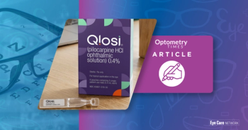
Orasis Pharmaceuticals, BlinkRx partnership offers new dispensing options for Qlosi
4
iVeena completes end-of-phase 1 FDA meeting for IVMED-85 in myopia
5