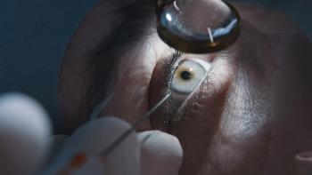
Sixth layer to human cornea discovered
A new layer of the human cornea has been discovered by Professor Harminder Dua, a researcher at the University of Nottingham in the UK.
A new layer of the human cornea has been discovered by a researcher at the University of Nottingham in the UK.
Professor Harminder Dua found the 15-micron thick layer between the corneal stroma and Descemet’s membrane. This makes the newly named Dua’s layer the fourth of six layers in the cornea. Dr. Dua shared his discovery in a recent study published in Ophthalmology. <http://www.aaojournal.org/article/S0161-6420(13)00020-1/abstract>
Dr. Dua suggests that this finding will affect corneal surgery, including penetrating keratoplasty, and understanding of corneal dystrophies and pathologies, such as acute hydrops.
This is an interesting finding that needs to be replicated by other labs in much younger eyes, according to Loretta Szczotka-Flynn, OD, PhD, professor of ophthalmology and visual sciences at Case Western Reserve University, andâ¨director of the contact lens service at University Hospitals Eye Institute in Cleveland.
“One possibility is that this may be an artifact occurring in older corneas because the only donor corneas they studied came from donors with a median age of 82 years,” Dr. Szczotka-Flynn says. “If the presence is confirmed, it will be very important in the lamellar types of corneal transplants. For example, in some lamellar transplants, such as DALK, surgeons may not want to bear all the way to Descemet’s. Rather, they may want to retrain their surgical techniques to bear down to this new proposed Dua’s layer, which will potentially keep the post-operative corneal transplant stronger and does not seem to lead to increased corneal haze.”
“This is an interesting read, but not without some controversy,” says Joseph P. Shovlin, OD, FAAO, private practitioner in Scranton, PA, and Optometry Times Editorial Advisory Board member. “Apparently, Binder et al described an acellular posterior stromal matrix layer in an original article in Investigative Ophthalmology and Vision Science.1 So, this may not be a novel (original) discussion of a sixth layer of the cornea. I am not certain of the clinical significance of this since we've had information on the structure for over a decade from Binder's paper.”
Dr. Binder’s paper states:
The attachment of Descemet’s membrane (DM) to the posterior stroma appeared to be accomplished in part by fibers 22.3 nm in diameter that ran perpendicular the DM. The depth of penetration of the fibers into DM was 0.16-0.21 um. They were associated frequently with a dense, amorphous mass at the interface between DM and the posterior stroma.1
Says Prof. Dua: “Perry Binder’s excellent work on the ultra structure of the cornea related to a different plane. The attachment of Descemet’s membrane to the Dua’s layer has also been studied by Prof. Friedrich Kruse, who refers to it as the inter-fascial layer and mentioned Binder’s paper.2”ODT
Reference
1. Binder PS, Rock ME, Schmidt KC, Anderson JA. High-voltage electron microscopy of normal human cornea. IOVS. 1991 July; 32: 2234-2243.
2. Schlötzer-Schrehardt U, Bachmann BO, Laaser K, Cursiefen C, Kruse FE. Characterization of the cleavage plane in Descemet’s membrane endothelial keratoplasty. Ophthalmology. 2011 Oct;118(10):1950-1957.
Newsletter
Want more insights like this? Subscribe to Optometry Times and get clinical pearls and practice tips delivered straight to your inbox.


