Treating the aging eye
In the aging eye, accommodation decreases; the crystalline lens yellows, hardens, and eventually opacifies; and systemic diseases such as arthritis, thyroid disease, cancer, diabetes, atherosclerosis, and high blood pressure take their toll on the eye. In addition, cognitive and functional limitations affect the aged.
Advances in medicine have extended the average life span of American men to 76.4 years and American women to 82.1 years,1 but greater life spans have brought one or more chronic illnesses to 80 percent of those over the age of 60.2
Along with their general medical problems, older patients must deal with declining vision and its physiological and psychological effects. Loss of vision can restrict one’s ability to carry out daily activities and lead to depression, social isolation, falls, fractures, and the inability to live independently.
In the aging eye, accommodation decreases; the crystalline lens yellows, hardens, and eventually opacifies; and systemic diseases such as arthritis, thyroid disease, cancer, diabetes, atherosclerosis, and high blood pressure take their toll on the eye. In addition, cognitive and functional limitations affect the aged.
They may not have support from their families or be unaware of available community services. Often changes in vision are undiagnosed and untreated. Patients may be living with unoperated cataracts, undiagnosed primary open-angle glaucoma, age-related macular degeneration, or diabetic retinopathy.
Keep in mind that one-third of new cases of blindness could have been prevented by early intervention.
Let’s look at some of the common visual conditions that affect our senior population.
Cataracts
Cataracts represent another common cause of visual loss in the elderly. Although we all will develop cataracts if we live long enough, the decrease in vision from cataracts is gradual, and not everyone who lives a normal life span will require surgery (see Figure 2).
In addition to age, causes of cataract include ultraviolet radiation from sunlight or other sources, corticosteroids, diabetes, family history, smoking, and previous eye injuries, inflammation, or surgery.6
As cataracts develop, the crystalline lens becomes yellow or cloudy. Initially, vision may be improved with a simple prescription change in eyeglasses.
As cataracts progress, they cause reduced visual acuity, increased glare, starbursts around headlights and streetlights at night, reduced color vision, and the need for more light when reading. These changes in vision are related to the size and location of the cataract and are generally slow and painless.
More from iTech:
Surgery becomes necessary when cataracts interfere with normal daily activities, such as driving, watching television, or reading the newspaper. Cataract surgery is the most frequently performed surgical procedure in the United States and has an excellent prognosis, with 90 percent of patients achieving vision of 20/40 or better.7
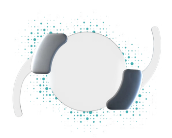
The surgery, a procedure called phacoemulsification, is done under local or topical anesthesia with IV sedation. A tiny incision is made, and the contents of the crystalline lens are emulsified, suctioned out, and replaced with an intraocular lens (IOL).
The IOL power is determined by presurgical measurements. We are now able to correct astigmatism with toric IOL designs and presbyopia with bifocal and multifocal IOL implants.
Macular degeneration
Age-related macular degeneration (ARMD) is a significant cause of vision loss in the elderly3 (see Figure 3). Risk factors include increasing age, family history, fair complexion and light irises, smoking, sleep apnea, metabolic syndrome (the most serious heart attack risk factors, including diabetes, prediabetes, abdominal obesity, high cholesterol, and high blood pressure), and high myopia.3
Initially, vision may be normal in spite of subtle degenerative changes, such as yellow, subretinal deposits known as drusen. In this “dry” form of ARMD, vision loss may be gradual. Straight lines may appear broken, wavy, or crooked, and patients may have difficulty reading or seeing road signs.
Macular degeneration can be demonstrated with the Amsler Grid (see Figure 1). Patients should wear their near correction when being tested. One eye is covered, and the chart positioned is 14 inches from the eye being tested. The patient is then asked to stare at the white dot in the center and notice if any of the lines on the grid appear to be wavy, broken, or missing.
More from iTech:
“Wet” ARMD usually starts out as the dry form and results in a sudden, significant loss of vision caused by leakage of blood or fluid from new, abnormally-formed vessels under the retina (subretinal neovascularization). Although it affects only about 20 percent of those who have macular degeneration, it accounts for two-thirds of the people with profound vision loss.4
ARMD affects only central vision. Patients develop a large central scotoma (blind spot), although they still maintain the ability to walk around without the assistance of a cane or seeing eye dog. Injections such as Eylea (aflibercept, Regeneron), Lucentis (ranibizumab, Genentech), and Avastin (bevacizumab, Genentech) may slow or stabilize vision loss by preventing the growth of leaky new blood vessels.5
Can we prevent the development of macular degeneration? Positive steps to take include stopping smoking, controlling cardiovascular disease, taking antioxidant dietary supplements, and following a diet high in fruits and vegetables, especially dark green, leafy vegetables like spinach and kale.
Glaucoma
Primary open-angle glaucoma, an optic neuropathy (optic nerve disease), is the second most common cause of visual loss among seniors.8 It causes changes in the optic nerve head, visual field loss, and in most cases, increased intraocular pressure (IOP), leading to blindness if left untreated. (See Figure 4.)
Risk factors include family history of glaucoma, high blood pressure, diabetes, myopia, African racial heritage, and elevated IOP.9 Early diagnosis and treatment can prevent optic nerve damage, visual field loss, and subsequent vision loss. Because pain is not associated with open-angle glaucoma, the disease may be well advanced, with significant visual field loss, before patients become aware of it.
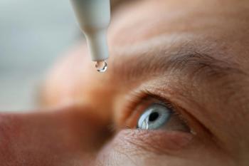
Many categories of medications are available to decrease intraocular pressure. Because seniors tend to be more sensitive to some glaucoma medications than younger patients and may also be taking systemic medications that can interact with their eye drops, the likelihood of side effects is greater in the elderly.
Side effects can be limited and systemic absorption reduced by covering the punctum (the tiny hole in the inner corner of the lower eyelid) and compressing the nasolacrimal duct when instilling eye drops. If IOP is not adequately controlled with eye drops, surgical intervention may be necessary.
Diabetic retinopathy
Diabetic retinopathy is the fourth most common cause of vision loss among the elderly in America.8 Over time, diabetes, especially poorly controlled diabetes, affects the circulatory system of the retina. Microaneurysms (tiny bulges that form and protrude from the walls of retinal blood vessels) can rupture and leak blood and fluids.
Symptoms are mild or nonexistent in the early stage, which is known as background or non-proliferative diabetic retinopathy, although leakage from the microaneurysms may cause macular edema (swelling and fluid retention).
As the disease progresses, new, fragile blood vessels form in the retina and vitreous (the gel that fills the back of the eye) and leak blood into the vitreous. This is known as proliferative diabetic retinopathy, which can cause severe vision loss and even blindness if left untreated.
Laser treatment is used to stop the leakage of blood and fluid and seal the abnormal, leaky blood vessels. It is important for the eyecare practitioner to work closely with the diabetic patient’s primary care physician or endocrinologist to maintain good control of blood sugar levels.
More from iTech:
Retinal occlusions
Total, sudden loss of vision may be caused by an embolus (blood clot or plaque) that lodges in and occludes the central retinal artery (central retinal artery occlusion). The loss of vision may be transient or permanent and requires immediate referral to an ophthalmologist.
The entire retina, except for the fovea (center of the macula), becomes edematous. Loss of a portion of the visual field can be caused by a branch retinal artery occlusion. In either case, treatment involves trying to move the embolus further downstream to minimize retinal damage, but loss of vision is often permanent.
Central or branch retinal vein occlusions can also occur and are caused by a thrombus (blood clot) blocking the vein that drains the blood from the eye. They are often seen in patients with high blood pressure, diabetes, glaucoma, and atherosclerosis, and require comanagement with the patient’s primary care physician.10
Temporal arteritis
Temporal arteritis, also known as giant-cell arteritis is an inflammation of the lining of the arteries that supply blood to the brain. Symptoms include head pain and tenderness, especially around the temples; scalp pain; jaw pain (claudication); sudden, permanent loss of vision in one eye; night sweats; and unexplained weight loss. Immediate referral to an ophthalmologist is critical to prevent loss of vision in the contralateral (opposite) eye. The condition is treated with steroids.
Dry eye syndrome
Dry eye syndrome, although a more benign condition, is still a significant problem among the senior population. Good tear quality and quantity is essential to maintain corneal integrity: to remove debris, to lubricate the eye, and to protect against disease. Keratitis sicca is the term used for markedly dry eyes.
Patient symptoms include burning, grittiness, excessive tearing, and injection (redness). Patients with rheumatoid arthritis and other collagen diseases may have been diagnosed with Sjögren’s syndrome, and live with dryness of the mouth and other mucus membranes in addition to dry eyes. Extreme dryness can lead to corneal damage and affect vision as well as comfort (see Figure 5).
In mild cases, artificial tears, used as needed, may provide sufficient relief. Restasis (cyclosporine A, Allergan) is a prescription eye drop that may increase tear production in patients whose tear deficiency is due to ocular inflammation associated with keratoconjunctivitis sicca (severe, chronic dry eye).11
Other dry eye treatments include punctal occlusion (silicone plugs placed in the tear drainage ducts to keep more tears in the eye), intense pulsed light therapy (IPL) that directs bursts of light at the lower eyelids and lower cheek areas to heat blocked eyelid glands; sleep masks that hydrate the eyes during the night; dry eye vitamins; and nutritional supplements such as flaxseed oil and fish oil.
Conclusion
Although the aging eye is affected by multiple conditions and diseases, technology and modern medicine enable eyecare practitioners and primary care physicians to work together and treat and manage many of them.
By making senior citizens aware of the importance of regular eye care, we can help them to benefit from new treatments and therapies, maintain their mobility and independence, and prevent the depression and social isolation that often occur when elderly patients are confronted with severe vision loss.
References:
1. Copeland L. Life expectancy in the USA hits a record high. USA Today. 2014 Oct 9. Available: http://www.usatoday.com/story/news/nation/2014/10/08/us-life-expectancy-hits-record-high/16874039/. Accessed 07/27/2015.
2. Council on Social Work Education. Chronic illness and aging. Available at: http://www.cswe.org/File.aspx?id=25462. Accessed 7/27/15.
3. National Eye Institute. Facts about age-related macular degeneration. Available at: https://nei.nih.gov/health/maculardegen/armd_facts. Accessed 07/27/2015.
4. American Society of Retina Specialists. Age-related macular degeneration. Available at: http://www.asrs.org/patients/retinal-diseases/2/agerelated-macular-degeneration. Accessed 7/27/15.
5. EyeSmart. Avastin, Eylea and Lucentis-What’s the difference? Available at: http://www.geteyesmart.org/eyesmart/living/avastin-eylea-lucentis-whats-the-difference.cfm. Accessed 07/27/2015.
6. Bailey G. Cataracts. AllAboutVision.com. Available at: http://www.allaboutvision.com/conditions/cataracts.htm. Accessed 07/27/2015.
7. Farzad F, Sarraf D, Coleman AL. Visual impairment in the elderly. Office Care Geriatrics. Ed. Rosental TC, Williams ME, Naughton BJ. Philadelphia: Lippincott Williams & Wilkins, 2006. 123. Print.
8. Quillen D. Common causes of vision loss in elderly patients. Am Fam Physician. 1999 Jul 1;60(1):99-108.
9. Mayo Clinic. Glaucoma: Risk factors. Available at: http://www.mayoclinic.org/diseases-conditions/glaucoma/basics/risk-factors/con-20024042. Accessed 07/27/2015.
10. Prevent Blindness. Central retinal vein occlusion. Available at: http://www.preventblindness.org/central-retinal-vein-occlusion. Accessed 7/27/15.
11. Allergan. Restasis prescribing information. Available here: http://www.allergan.com/assets/pdf/restasis_pi.pdf. Accessed 07/27/2015.
Newsletter
Want more insights like this? Subscribe to Optometry Times and get clinical pearls and practice tips delivered straight to your inbox.


