When corneas go viral: Epidemic keratoconjunctivitis edition
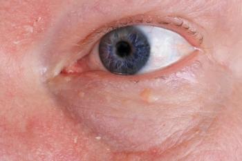
The possibility that a given garden-variety case of viral conjunctivitis is a burgeoning epidemic keratoconjunctivitis (EKC) is enough to intimidate any physician.
The possibility that a given garden-variety case of viral conjunctivitis is a burgeoning epidemic keratoconjunctivitis (EKC) is enough to intimidate any physician.
The term “pink eye” is a broad term often used to describe conjunctivitis caused by both bacteria and a virus; however, in this article we will refer exclusively to viral conjunctivitis. Its contagious nature universally strikes dread into both practitioners and patients alike.
It is with good reason that we approach pink eye with trepidation-it is estimated that up to 45 percent of people in an EKC patient’s close surroundings may become infected.1 In this article we will discuss transmission, diagnosis, ocular and corneal manifestations as well as current and future thoughts regarding treatment.
Virus transmission
Since 1953, seven species (named A through G) and 54 serotypes have been named for the systemic adenovirus.2 As a whole, these serotypes are known to cause infections ranging from gastroenteritis to respiratory tract infections to several varieties of viral conjunctivitis.
At least 19 out of the 54 serotypes are known to cause eye infection-those most commonly associated with causing EKC are adenovirus 8, 19, and 37.1 Interestingly, less than 5 percent of the general U.S. population has immunity to type 8 antibodies, so our fears of low natural immunity seem to be founded because almost every individual is considered susceptible.1
EKC’s incubation period is between 2 and 14 days, during which time patients may not be aware they are carrying the infection. Patients may remain infectious for over 14 days after the symptoms begin.1,2 It is during the symptomatic period when “viral shedding” occurs-manifested by the classic watery and crusty eyes. Infectious cell lysis into the ocular fluids and onto unsuspecting objects and individuals is usually transmitted via direct contact with the eyes by fingers or any other “carrier” object.2
Related:
Environmental contaminants are so highly contagious that the Centers for Disease Control and Prevention (CDC) published a Morbidity and Mortality Weekly Report in 2013 detailing six unrelated EKC outbreaks from 2008 to 2010 in Florida, Illinois, Minnesota, and New Jersey, totaling over 400 total patients.3
As frontline examiners of EKC, how can we prevent transmission?
First and foremost is triaging suspected cases to isolated areas, preventing patients from spreading the infection in the waiting room to pens, clipboards, doorknobs, and the like. Handwashing by the clinician, or better yet, donning disposable gloves is imperative. Cleaning instruments with buffered bleach solution (not alcohol swabs) and allowing to fully dry helps break down the virus.
Diagnosing EKC
Diagnosis of EKC is via one of two ways: laboratory detection and clinical examination.
If practicing in or near a large hospital setting, one method to consider is the laboratory route. Cell culture with confirmatory immunofluorescence assay (CC-IFA) visualizes adenovirus particles by allowing them to replicate and produce viral progeny-positive test results mean the patient has the virus and is contagious.2
Another method is by polymerase chain reaction (PCR), which is able to measure not only live and infectious adenovirus, but also partial or dead virus. PCR shows better total quantity of virus load.
Related:
The newest method of detection-easy to complete in-office by any practitioner-is AdenoPlus (Rapid Pathogen Screening), a 10-minute, painless, and fairly accurate detection test.2 The sensitivity of AdenoPlus is 89 percent, and its specificity is 94 percent.4 To perform the test, a small sterile strip is used to collect tears from the patient’s eye and then dipped into a buffer solution. Similar to a pregnancy test, the buffer leaves a single line mark (signifying negative) or a double-line mark (signifying positive).
Diagnosis via ocular manifestations is the mainstay of our practices. Patient complaints range from burning and itching eyes to watery discharge to a red eye and/or having it “glued shut.” Patients may even mention discomfort anterior to the ear, signifying a swollen preauricular lymph node (PAN) and may present with swollen lids. It is important to examine the patient for preauricular lymphadenopathy because about one-third to one-half of adenovirus cases has concomitant nodes with follicles-both are part of the same lymphogenesis.2 Examine by gentle palpation anterior to the tragus of the ear, which the patient may report is tender.
Careful examination of the palpebral conjunctiva often reveals follicles and rubeosis of both the inferior and superior conjunctiva. Everting the upper lid, if possible, may show large follicles sometimes verging on giant. (See Figures 1 and 2.)
Manifestations
Corneal manifestations may bring with them more patient visual complaints. Keratitis occurs in approximately 80 percent of EKC patients and can cause increased tearing, photophobia, blurry vision, and foreign body sensation.2 Following the keratitis, over 40 infiltrates can develop (usually around Day 11), but are most prominent in Weeks Three and Four.2 It is estimated that between 30 and 50 percent of patients with EKC will develop these infiltrates, which may cause persistent clouding of the vision and increased light sensitivity.2 Some corneas will develop only a few infiltrates that fluctuate in activity-some lying dormant, and some remaining inflamed (Figure 3).
EKC-related infiltrates have been known to reactivate sometimes up to three years post-initial infection, not to the severity of the first episode but enough that they often cause symptoms meriting intervention. During recurrent episodes, the bulbar conjunctiva of these eyes may have some inflammation, and there may be some follicular reaction but again not to the severity of the initial attack. Even after complete inactivity of the infiltrates, sites of previous infiltrates may remain permanently scarred, and depending on location, continue to affect a patient’s vision.
Related:
Treating EKC
Treatment of EKC can be frustrating and slow going. There is currently no commercially available treatment for adenovirus, so treatment regimens usually target the symptoms and auxiliary effects of the virus.
In the cornea’s reaction to adenovirus, symptoms like irritation, watering, photophobia, pain, foreign body sensation, eyelid swelling, and pain may accelerate a practitioner’s decision for treatment. In these cases, topical anti-inflammatories and antibiotic and sometimes anti-immune therapies may be required.
In terms of anti-inflammatories, topical steroids are widely, and sometimes controversially, used.1 It is generally believed that steroids, while they may resolve some acute symptoms of irritation, may actually prolong the persistence of the viral shedding, replication, and infection in the cornea.1,2
However, when corneal infiltrates are present, intervention to treat the active infection may require a course of a mild steroid, such as fluorometholone (FML, Allergan) or loteprednol etabonate (Lotemax, Bausch + Lomb). The dosage of these usually begins at four times per day and requires a long tapering process over weeks, rarely over months.
When corneal manifestations require anti-inflammatory utilization, sometimes the most acute phase of infiltrates may merit from a pulse dose of antibiotics. Though the antibiotics will have no effect on the viral replication nor shedding if infiltrates have caused large enough epithelial loss, bacterial coverage could prevent a concurrent infection. In addition, in the event of pseudomembrane growth and removal, antibiotics prevent superinfection of the delicate palpebral conjunctiva.
There is active discussion on the taper schedule of topical steroid use for infiltrates that persist long past the follicular viral episode. In some cases, infiltrates may persist even two years after the initial infection, causing various levels of inflammation and symptoms. After a prolonged taper of steroids, potentially over many months, patients’ corneas may be quiet for some time. In a small percentage of these, the infiltrates may recur again-never to the initial degree of severity- but significant enough to bring the patient back into our chair.
Related:
The discussion then focuses on what to do. Begin another course of steroids at a low dose and taper again? Long-term steroid use-even at low concentrations and dosages-may still lead to unwanted side effects such as cataract and glaucoma in addition to dependence and irritation. Some advocate the use of anti-immune therapy with cyclosporine A (Restasis, Allergan).5 Topical CsA plays a role in suppressing the immune reaction of the cornea and may decrease the activity and persistence of remaining or recurrence of infiltrates.
The newer treatment avenue-still in development as of 2011 for treatment of adenoviral conjunctivitis-is a povidone-iodine 0.4% and dexamethasone 0.1% ophthalmic suspension.1,6 Similar in thought to the still-employed Betadine (povidone-iodine, Purdue Products) rinse in hopes of prevention of adenoviral proliferation, this topical approach dually provides antimicrobial activity and may reduce viral shedding.2 The potential to lessen severity and prevent communication would be a fantastic addition to our current arsenal.
Epidemic keratoconjunctivitis, in addition to its fearful contagious potential, can also be excruciatingly difficult to treat. Early recognition by the practitioner and prevention of spread to the other eye and other individuals is vitally important in curtailing this infection. Acute intervention upon corneal involvement may be necessary with regular follow-up visits and refinement of the treatment regimen.
References
1. Adenovir Pharma. About epidemic keratoconjunctivitis (EKC) – a serious disease without effective treatment. Available at:
2. Abelson MB, Shapiro A. A guide to understanding adenovirus, the diseases it causes and the best ways to treat these conditions. Rev Ophthalmol. Available at:
3. Centers for Disease Control and Prevention. Adenovirus-Associated Epidemic Keratoconjunctivitis Outbreaks-Four States, 2008–2010. MMWR. 2013 Aug;62(32):637-641.
4. Sambursky R, Tauber S, Schirra F, Kozich K, Davidson R, Cohen EJ. The RPS adeno detector for diagnosing adenoviral conjunctivitis. Ophthalmology. 2006 Oct;113(10):1758-64.
5. Okumus S, Coskun E, Tatar MG, Kaydu E, Yayuspayi R, Comez A, Erbagci I, Gurler B. Cyclosporine a 0.05% eye drops for the treatment of subepithelial infiltrates after epidemic keratoconjunctivitis. BMC Ophthalmol. 2012 Aug 18;12:42.
6. Clement C, Capriotti JA, Kumar M, Hobden JA, Foster TP, Bhattacharjee PS, Thompson HW, Mahmud R, Liang B, Hill JM. Clinical and antiviral efficacy of an ophthalmic formulation of dexamethasone povidone-iodine in a rabbit model of adenoviral keratoconjunctivitis. Invest Ophthalmol Vis Sci. 2011 Jan 21;52(1):339-44.
Newsletter
Want more insights like this? Subscribe to Optometry Times and get clinical pearls and practice tips delivered straight to your inbox.


