

|Articles|October 9, 2020
- October digital edition 2020
- Volume 12
- Issue 10
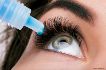
Where to begin with dry eye technology
Author(s)Eric Botts, OD
Adding laboratory testing and meibography helps patients and practices
Advertisement
Most ODs utilize slit-lamp evaluation, lissamine green staining, and phenol red thread testing for dry eye diagnosis. Additional instrumentation, including in-office lab tests, meibography, and intense pulsed light provides better monitoring and therapy for dry eye patients. New technology gives ODs an edge in achieving tear film homeostasis.
Dry eye diagnosis and treatment is quickly becoming one of the fastest ways to increase patient volume and collected fees in optometric practices. More and more doctors are adding dry eye clinics successfully to their practices because dry eye disease is quickly increasing in numbers primarily because the new technology utilized to diagnose it has improved significantly.
Although a thorough slit-lamp evaluation, lissamine green staining, and phenol red thread testing is a good start for diagnosing dry eye disease, I encourage ODs to add additional dry eye testing to their arsenals of clinical tools.
In-office lab tests
Start with two laboratory tests that will improve ODs’ ability and confidence in accurately determining which patients require dry eye therapy.
TearLab Osmolarity System measures the osmolarity of the tear film in a repeatable manner that provides practitioners with a number to determine whether the patient’s tear osmolarity is normal, minimally dry, moderately dry, or severely dry. My patients want to know their TearLab numbers, and knowing numbers improves treatment compliance when I can show patients the positive effect of their follow through with the treatment plans I have provided them.
InflammaDry (Quidel) is another test that measures inflammation on the ocular surface. Inflammadry results guide my decision on whether to insert punctal plugs as part of dry eye therapy. A positive Inflammadry result typically requires topical treatment with anti-inflammatory medication prior to insertion of punctal plugs, whereas a negative result provides an immediate opportunity to insert punctal plugs to deliver dry eye therapy.
TearLab and Inflammadry can be easily performed by technicians, allowing the doctor to quickly review results with the patient. Both TearLab Current Procedural Terminology (CPT) 83861 and Inflammadry CPT 83516 are billable procedures to medical insurance plans that require a Clinical Laboratory Improvement Amendment (CLIA) waiver. Submit both of these laboratory tests using a QW modifier to receive full reimbursement from most medical insurance carriers. Apply for a waiver by completing an application available on the Centers for Medicare & Medicaid Services (CMS) CLIA website at CMS.gov or from the local state Department of Health agency.
Both of these laboratory tests address the latest definition for dry eye based on the Tear Film and Ocular Surface Society’s (TFOS) Dry Eye Work Shop (DEWS) ll definition: Dry eye is a multifactorial disease of the ocular surface characterized by a loss of homeostasis of the tear film, and accompanied by ocular symptoms, in which tear film instability and hyperosmolarity, ocular surface inflammation and damage, and neurosensory abnormalities play etiological roles.1
Meibography
Meibography is an another test I frequently perform to diagnose and monitor dry eye disease. This test may be performed with any number of instruments available, including the Keratograph 5M (Oculus), CA-800 Corneal Analyzer (Topcon), LipiVew/LipiScan (Johnson & Johnson Vision), and Meibox (Box Medical Solutions).
Meibography is an integral part of diagnosing dry eye disease because it shows the meibomian gland function on both inferior and superior lids. Meibography is an objective test that is also a very useful education tool to show the patient how her meibomian gland dysfunction may contribute to her chronic dry eye disease.
Although meibography is not a billable procedure, external photos are reimbursable, and often I order both tests and am reimbursed for the external photos (CPT 92285).
In today’s busy practice, space for additional instrumentation has become an obstacle for many ODs. Fortunately, dry eye laboratory testing has a very small footprint. Plus, most of the meibography units will also perform corneal topography and exterior photos so ODs can have the benefit of multiple tests performed by one instrument.
Many patients today are being treated with thermal devices combined with physical expression of the meibomian glands. Several procedures and instruments available today include:
– LipiFlow (Johnson & Johnson Vision)
– iLux (Alcon)
– TearCare (Sight Sciences)
– MiBo Thermoflo (MiBo Medical Group)
Intense pulsed light
Intense pulsed light (IPL) therapy also provides treatment for dry eye disease that involves using light energy to liquefy and release oil from clogged meibomian glands.
Important to note is that none of the above thermal treatments or IPL are currently reimbursed by any medical insurance carriers, resulting in all procedures being out-of-pocket expenses for patients. ODs should evaluate the return on investment before purchase of any equipment to insure it is the right instrument for your practice.
Making it work
I have reviewed my practice treatment protocol for dry eye disease to make my examinations as streamlined and efficient as possible. My protocol for dry eye allows most diagnostic tests, including laboratory tests and meibography, to be performed before I see the patient. This allows me to focus on reviewing test results and determining the best treatment plan for my patient.
Most of my dry eye patients are previous patients whose prior complaints and symptoms were not enough to elicit a diagnosis of dry eye. However, with the addition of TearLab, InflammaDry, and meibography, these patients are no longer slipping through the cracks undiagnosed.
See box for potential practice revenue by incorporating dry eye diagnosis and treatment to an optometric practice.
Investing in a dry eye clinic for the practice offers more than just a financial return—it provides better care for patients. Many patients suffering from dry eye symptoms are tolerating their conditions because it is not accurately diagnosed.
ODs already have the clinical skill set to diagnose and treat dry eye, so the only question is are they willing to invest in the instrumentation and laboratory tests necessary to efficiently detect dry eye and treat it?
Dr. Botts earned his Doctorate of Optometry from Southern College of Optometry and has served as past president of the Illinois Optometric Association and Energeyes. [email protected]
Reference
1. Craig JP, Nichols KK, Akpek EK, Caffery B, Dua HS, Choun-Ki J, Liu Z, Nelson JD, Nichols JJ, Tsubota K, Stapleton F. TFOS DEWS II definition and classification report. Ocul Surf. 2017 Jul;15(3):276-83
Articles in this issue
over 5 years ago
4 vision-related causes of contact lens discomfortover 5 years ago
Which patients are poor laser vision correction candidates?over 5 years ago
AI and the modern optometric practice restartover 5 years ago
When a retinal detachment isn’t a retinal detachmentover 5 years ago
Pipeline: Potential new therapy coming for macular diseasealmost 6 years ago
What I have learned from the COVID-19 pandemicAdvertisement
Related Content
Advertisement


Latest CME


Advertisement
Advertisement
Trending on Optometry Times - Clinical News & Expert Optometrist Insights
1
AOA 2026: AREDS2 and AREDS2+B vitamin complex implications for AMD
2
AOA 2026: The latest updates in keratoconus treatment advancements
3
Thea Pharma launches Zolymbus in the US as preservative-free bimatoprost gel
4
First EDOF contact lens improves intermediate, near visual acuity in presbyopes, study finds
5