
|Articles|August 27, 2020
Why lid wiper epitheliopathy is a game changer
Lid wiper epitheliopathy is a critical clinical marker that will not only help identify an abnormality in ocular surface homeostasis, but also a key clinical marker to track when treating a patient for dry eye
Advertisement
Homeostasis is critical when it comes to a healthy ocular surface. ODs are on a constant quest to improve diagnostic and therapeutic capabilities in order to maintain or restore ocular surface homeostasis. In the relentless quest to pursue this, appropriately identifying the absence of homeostasis is of increasing importance. ODs know that an ocular surface that does not maintain homeostasis will affect vision, ocular comfort, and contact lens comfort.
The evolution of diagnostics in the realm of ocular surface disease has advanced tremendously. For those offices that have advanced diagnostics for ocular surface disease, we are certain that it has changed the way ODs think of the ocular surface. But don’t forget about some of the basic tools that we all have access to that can provide us a better understanding of the ocular surface and the presence or absence of homeostasis.
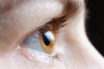
The lid wiper area
Vital dyes are critical in order to help understand the way that the ocular surface functions. One key anatomical structure that is often overlooked but is certainly something that is important to understand with respect to ocular surface disease is the lid wiper area (LWA). The LWA is the small area that resides posterior to the meibomian glands and is responsible for the wiping action of the upper eyelid along the ocular surface during the blink. Its anterior portion is marked by the Line of Marx (LOM).
The LOM is a critical ocular structure that is the junction between the anterior keratinized epithelium of the lid and the posterior mucous membrane on the palpebral conjunctiva.1 Under normal circumstances the LOM resides posterior to the meibomian gland orifices. In times of inflammation or long-term ocular surface abnormality, the LOM can migrate anteriorly and it can also become irregular.2 This is a critical landmark to look for when assessing the ocular surface. The LOM stains with rose bengal, lissamine green, and fluorescein and can be easily visualized with either one of these dyes.
The LWA can be incredibly telling of the level of friction that exists between the ocular surface and portion of the upper eyelid that wipes along the ocular surface during every blink. Remember that the upper eyelid constitutes most of the blink in a normal individual. As such, it is absolutely critical that the ocular surface including the tear film is functioning appropriately in order to provide appropriate lubrication between the LWA and the ocular surface.
If the tear film is not functioning appropriately, it increases the level of friction between the LWA and the ocular surface that it “wipes” over. This excessive friction can irritate the epithelium, resulting in staining that manifests itself on the LWA, termed lid wiper epitheliopathy (LWE).3Rose bengal, lissamine green, and fluorescein will all be absorbed by the tissue if LWE is present.
The evaluation of the LWA has to be a conscious decision by the practitioner because it requires vital dyes being placed on the ocular surface and eversion of the upper lid. We feel it is a critical measurement as an early indicator of an abnormal relationship between the LWA and the ocular surface represented by reduced lubrication secondary to an insufficient tear film.
Several papers have demonstrated the importance of the presence of LWE and dry eye symptoms. Korb et al demonstrated that of patients reporting symptoms of dry eye, 76 percent had LWE.3 In another study, Korb et al found that of those patients complaining of contact lens discomfort, 80 percent had LWE.4
It is critical to understand how to metricize LWE in order to determine what level of LWE ODs are seeing and whether treatment is influencing the extent of LWE. It is a combination of horizontal length and the sagittal width of the region of staining present. If the horizontal length is less than 2 mm, a score of 0 appears. If it is 2 to 4 mm, a score of 1 will be given. If it is 5 to 9mm, a score of 2 is given, and if it is 10 mm or greater, it is given a score of 3.
The sagittal width is an estimate of the percentage of the LWA that is occupied by stain or LWE. If it is less than 25 percent, the LWE score is 0.If it is 25 to 49 percent it is 1, if it is 50 to 74 percent it is 2 and if it is 75 percent or greater, it is given a score of 3. The total LWE score is simply an average of the score of the horizontal length and the sagittal width. So, if the horizontal length was a 3 and the sagittal width score was a 1, the final LWE score is 2.
Lid wiper epitheliopathy is a critical clinical marker that will not only help identify an abnormality in ocular surface homeostasis, but also a key clinical marker to track when treating a patient for dry eye. Keep this in mind while managing ocular surface disease, and ODs will be enlightened with what they see.
References
1. Doughty MJ, Naase T, Donald C, Hamilton L, Button NF. Visualisation of “Marx's line” along the marginal eyelid conjunctiva of human subjects with lissamine green dye. Ophthalmic Physiol Opt. 2004 Jan;24(1):1-7.
2. Yamaguchi M, Kutsuna M, Uno T, Zheng X, Kodama T, Ohashi Y. Marx line: fluorescein staining line on the inner lid as indicator of meibomian gland function. Am J Ophthalmol. 2006 Apr;141(4):669-675.
3. Korb DR, Herman JP, Greiner JV, Scaffidi RC, Finnemore VM, Exford JM, Blackie CA, Douglass T. Lid wiper epitheliopathy and dry eye symptoms. Eye Contact Lens. 2005 Jan;31(1):2-8.
4. Korb DR, Greiner JV, Herman JP, Hebert E, Finnemore VM, Exford JM, Glonek T, Olson MC. Lid-wiper epitheliopathy and dry-eye symptoms in contact lens wearers. CLAO J. 2002 Oct;28(4):211-216.
Advertisement
Related Content
Advertisement


Latest CME


Advertisement
Advertisement
Trending on Optometry Times - Clinical News & Expert Optometrist Insights
1
Johnson & Johnson invests $1B in ACUVUE contact lens manufacturing
2
Self-screening for ocular surface malignancies using a smartphone
3
Epion Therapeutics completes phase 3 trials of EpiSmart epithelium-on cross-linking for keratoconus
4
Johnson & Johnson expands ACUVUE OASYS 1-Day for Astigmatism parameter range
5