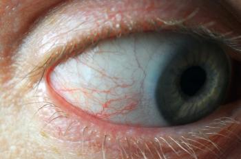
A closer look at central retinal artery occlusion
A central retinal artery occlusion (CRAO) represents a true ocular emergency. It is frequently caused by an embolus originating from carotid artery disease secondary to atherosclerotic plaques.
A central retinal artery occlusion (CRAO) represents a true ocular emergency. It is frequently caused by an embolus originating from carotid artery disease secondary to atherosclerotic plaques. The embolus can also originate from a cardiac source. There is a poor visual prognosis for a CRAO because there is currently no effective treatment to restore vision in the affected eye.
History
A 63-year-old white male presented to the emergency room (ER) complaining of sudden vision loss in the left eye two days prior and was referred to the eye clinic for evaluation. The patient denied any symptoms of jaw claudication, sensitivity to the temporal area, flashes, floaters, pain, paresthesia, numbness, headache, or neurological deficits. The patient also denied any ocular trauma or previous history of ocular disease. The patient reported that “bits and pieces” of his vision were clear, but his superior field of vision was obscured. He could see only a “strip of vision” out of his left eye.
Related:
The patient’s blood pressure in the ER that day was 187/114 mm Hg. His medical history was positive for type II diabetes mellitus and coronary artery disease. His cardiac history included bypass surgery and stenting. The patient’s blood glucose measured 134 mg/dL that day, and his last documented HbA1c was 7.7 six months ago.
Diagnostic data
The patient’s entering corrected visual acuity at distance was 20/30+ OD using the
Snellen chart and 6/200+ OS using the Feinbloom chart. Pupils were reactive to light, and an afferent pupillary defect was present in the left eye. Confrontation visual fields were full to finger counting OD and constricted 360 degrees OS. Intraocular pressure (IOP) measured 19 mm Hg OD and 12 mm Hg OS. Slit lamp examination revealed trace injection of the bulbar conjunctiva. Both corneas were clear, the anterior chambers were free of cells and flare, and there was no neovascularization of the iris.
Related:
Fundoscopy revealed nuclear and posterior subcapsular cataracts in both eyes as well as posterior vitreous detachments OU. The cup-to-disc ratio was 0.4/0.35 OD and 0.5/0.45 OS with no evidence of swelling, edema, or hemorrhages of the optic nerve in either eye.
The posterior pole of the right eye was positive for one Hollenhorst plaque inferior-temporal to the fovea. The posterior pole of the left eye contained about 15 to 20 calcium fibrous and Hollenhorst plaques. The majority of the plaques were located at bifurcations.
The macula of the left eye contained a cherry red spot, and the retina was pale. There was also one blot heme associated with a calcium plaque at an arteriole bifurcation nasal in the midperipheral retina in the left eye. The peripheral retina was clear of lattice or retinal breaks OU. (See Figures 1 and 2.)
Amsler grid testing revealed a central and nasal scotoma in the left eye. With color vision testing using Ishihara plates, the patient could distinguish 3/14 pseudoisochromatic plates OD and only the test plate using eccentric fixation OS. The patient later reported a longstanding history of a color deficiency.
Diagnosis
Based upon the clinical findings, the patient was diagnosed with a CRAO in the left eye with cholesterol, calcific, and fibrous plaques present.
Treatment and follow-up
Because of the strong association between a CRAO and systemic disease, the patient was referred immediately to his primary care provider (PCP) and cardiologist for further evaluation and systemic treatment. The PCP requested an echocardiogram and considered changing the patient’s anticoagulant treatment; the patient was currently on 325 mg of aspirin (ASA). The patient was scheduled to return to the retina clinic in three weeks for follow-up.
Related:
Results of an echocardiogram and carotid Doppler showed complete occlusion of the left internal carotid artery and stenosis in the lower range of 60 to 79 percent of the right internal carotid. An MRI with contrast also revealed marked atherosclerotic changes of the left vertebral artery, probable mild stenosis of the right subclavian artery, and possible severe stenosis of the distal basilar artery.
Magnetic resonance angiogram (MRA) of the head/neck confirmed a complete occlusion of the left internal carotid artery (ICA), 64 to 68 percent stenosis of the origin of the right ICA, and severe stenosis of the origin of the right external carotid artery.
In addition, there was marked atherosclerotic changes of the non-dominant left vertebral artery and probable mild stenosis of the origin of the right subclavian artery. As a result of these findings, the patient’s PCP initiated treatment with Aggrenox (aspirin/extended-release dipyridamole, Boehringer Ingelheim) and changed the ASA dosage to 81 mg.
At the follow-up visit in the retina clinic three weeks later, visual acuity measured 20/25+2 OD and CF OS. A dilated fundus exam revealed the same clinical findings as the initial visit. The plan at this point was to continue to follow up with the patient’s PCP and the vascular clinic and return in two months for a dilated fundus exam.
The patient’s PCP recommended continuation of the more aggressive antiplatelet treatment and regular surveillance with carotid duplex ultrasound. If new symptoms developed involving the right hemisphere, then surgery would be indicated.
One year later, MRA of the neck showed marked progression of the right ICA stenosis of at least 84 percent. The patient would go on to have neurological symptoms and underwent a right carotid endarterectomy through a private hospital.
Discussion
Retinal arteriolar emboli have been associated with a higher risk of cardiovascular disease and stroke and have a prevalence of about 1.4 percent in people over the age of 40.1 Retinal arteriolar plaques may appear as white and glistening.
These crystalline particles, known as Hollenhorst plaques (HHP), tend to lodge at arteriolar bifurcations. They are thought to originate from the inner layers of carotid atheromas and represent cholesterol deposits. They may rarely cause transient vision loss but more often will fragment and pass through the retinal circulation without incident. Solid, white, nonrefractile plaques that lodge in the larger vessels around the optic nerve head originate from calcification of cardiac valves.
These calcific emboli are often occlusive and may cause retinal infarcts. Long dull-white plugs represent platelet emboli from the surface of atheromas. They are mobile and will undergo fragmentation and dissolution but can cause transient obstruction to retinal blood flow. Transient visual loss (TVL) may occur. Linear white deposits within arterioles probably represent fibrin or thromboembolic material from the walls of the heart. They may result in retinal artery occlusions and subsequent infarcts.2
CRAO has commonly been described as being caused by an embolism from the carotid artery bifurcation.3 More specifically, it is believed that the origin of the CRAO is an embolus from the cardiovascular system that travels through the ophthalmic artery to the central retinal artery and lodges at the lamina cribrosa.4
The condition typically presents as a painless, unilateral loss of vision. Clinical manifestations of an acute CRAO include a cherry red spot in the macula with a pale retina.3,4 The macular cherry red spot is due to the presence of choroidal circulation.4
Management of a CRAO revolves around management of the systemic disease; it has been estimated that there is a 20 percent mortality rate within five years of the diagnosis.5 Work-up includes an immediate erythrocyte sedimentation rate (ESR) to rule out giant cell arteritis (GCA) if the patient is older than 55 years of age.
If the patient’s history and/or ESR are consistent with GCA, high-dose systemic steroids are started. Other blood tests include: fasting blood sugar (FBS), complete blood count (CBC) with differential and platelets, and lipid profile. In addition, a carotid artery evaluation is performed (Doppler and ultrasound of the carotid arteries) as well as a cardiac evaluation (echocardiogram and Holter monitor).6
Related:
The detection of emboli as an initial presentation of an underlying systemic disease may afford the opportunity for early intervention. Two major prospective studies, North American Symptomatic Carotid Endarterectomy Trial (NASCET) and the European Carotid Surgery Trial (ECST) provide strong evidence for the benefit of carotid endarterectomy in symptomatic patients when performed by experienced surgeons.
Both studies showed that in patients with retinal or hemispheric symptoms attributed to severe (70 to 99 percent) carotid stenosis, endarterectomy was superior to medical care alone. There was no benefit of carotid endarterectomy in symptomatic patients with mild stenosis (less than 50 percent).7
Patients with carotid artery disease, who are asymptomatic, are prevalent in the general population.
However, severe (greater than 70 percent) carotid stenosis of the asymptomatic patient is rare in comparison with symptomatic patients. Four randomized clinical trials were conducted (Carotid Artery Surgery Asymptomatic Narrowing Operation Versus Aspirin [CASANOVA], Mayo Asymptomatic Carotid Endarterectomy [MACE] Trial, Veterans Affairs Asymptomatic Carotid Endarterectomy Trial, and the Asymptomatic Carotid Atherosclerosis Study [ACAS]) to determine the risks and benefits of carotid endarterectomy in asymptomatic patients.
CASANOVA, MACE, and the Veteran Affairs Asymptomatic Trial found no benefit from surgery in asymptomatic patients.
Based on a five-year projection, the ACAS found that carotid endarterectomy reduced the risk of absolute stroke by 5.9 percent and the relative risk of stroke and death by 53 percent. Patients enrolled in ACAS were younger than 80 years and had asymptomatic carotid stenosis of 60 percent or more.7
A retrospective review of incidental retinal emboli found on diabetic retinopathy screenings recommended that alerting a patient’s PCP of retinal arteriolar findings can help identify patients with vascular disease who can benefit from antiplatelet therapy. Although there are no current strategies to distinguish who can benefit most from additional testing, the review recommended referral to the PCP for possible initiation of treatment; in the review, 28 percent of patients required medical interventions such as antiplatelet therapy while three percent required surgical intervention.1
One study suggested that the internal carotid artery ipsilateral to the affected eye had a significantly higher incidence of severe carotid artery stenosis compared to the non-affected side and played an important role in retinal ischemia.3
Another study determined that about nine percent of patients with an asymptomatic HHP had significant carotid stenosis (stenosis greater than 60 percent); the study suggested that patients with HHP but no symptoms of visual change may not necessarily require routine screening unless there are other signs of carotid stenosis. Carotid duplex scanning is indicated to investigate the level of carotid stenosis.8
Yet another study investigated whether ocular findings justified further testing, specifically carotid imaging.
The findings showed that 18.2 percent of patients with Hollenhorst plaques had significant carotid stenosis (defined as greater than 60 percent).9
The study calculated that the presence of HHP has a 50 percent sensitivity for being an indicator of significant carotid occlusive disease. Interestingly enough, the study suggested that retinal artery occlusion did not display significant carotid stenosis (greater than 60 percent).10
One particular study tried to examine the association between CRAO, BRAO, and HHP and the risk of cerebrovascular events and death in an ethnically diverse population. It concluded that the presence of a CRAO or BRAO is associated with a higher rate of cerebrovascular events and death, whereas the finding of HHP does not appear to increase the risk of either.
These results suggest that an evaluation, including cost-utility analysis, of current clinical practice following the identification of HHP may be warranted.11
There is no current treatment to significantly improve visual prognosis after suffering from a CRAO. There is a retrospective study that suggests that intra-arterial thrombolysis may improve visual outcomes, especially for younger patients who present within four hours of the onset of visual symptoms; complications can include transient ischemic attacks and stroke.12
However, the current focus is to treat the underlying systemic condition and preserve the vision in the unaffected eye.
References:
1. Ahmed R, Khetpal V, Merin LM, et al. Case Series: Retrospective review of incidental retinal emboli found on diabetic retinopathy screening: Is there a benefit to referral for work-up and possible management? Clin Diabetes. 2008 Oct 1;26(4):179.
2. Marks ES, Adamczyk DT, Thomann KH. Primary Eyecare in Systemic Disease. Connecticut: Appleton & Lange, 1995.
3. Kimura K, Hasimoto Y, Ohno H, et al. Carotid artery disease in patients with retinal artery occlusion. Intern Med. 1996 Dec; 35(12):937-40.
4. Alexander LJ. Primary Care of the Posterior Segment. 3rd edition. New York: McGraw Hill, 2002.
5. Wong TY. The Ophthalmology Examinations Review. 2nd Edition. Singapore: World Scientific, 2011
6. Cullom RD, Chang B. The Wills Eye Manual. 2nd Edition. Philadelphia: J.B. Lippincott Company, 1994.
7. Trego ME, Pagani JM. Three presentations of monocular vision loss. Optometry. 2006 Feb;77(2):82-7.
8. Bruce AS. Posterior Eye Disease and Glaucoma A-Z. Edinburgh: Elsevier, 2008.
9. Friedman NJ. The Massachusetts Eye and Ear Infirmary Illustrated Manual Ophthalmology. 3rd edition. Saunders: Elsevier, 2009.
10. McCullough HK, Reinert CG, Hynan LS, et al. Ocular findings as predictors of carotid occlusive disease: Is carotid imaging justified? J Vasc Surg. 2004 Aug;40(2); 279-86.
11. Giacovelli JK, Mozayan A, Mian U. Retinal artery occlusions, Hollenhorst plaques and cerebrovascular events and mortality in the Bronx. Invest Ophthalmol Vis Sci. ARVO Meeting Abstracts March 26, 2012. 53:988.
12. Arnold M. Comparison of intra-arterial thrombolysis with conventional treatment in patients with acute central retinal artery occlusion. J Neurol Neurosurg Psychiatry. 2005 Feb;76(2); 196-9.
Newsletter
Want more insights like this? Subscribe to Optometry Times and get clinical pearls and practice tips delivered straight to your inbox.


