AAOpt 2025: Diabetes case studies with an eye to the future with Dr Paul Chous
How can optometrists leverage new technologies to improve early detection, monitoring, and management of diabetic eye conditions? This question and more concerning the care for patients with diabetes were tackled by A. Paul Chous, MA, OD, FAAO and Julie Rodman, OD, MSc, FAAO, FORS during their presentation given at the American Academy of Optometry (AAOpt) Academy meeting in Boston Massachusetts. In an exclusive interview with Optometry Times, Chous outlined what the future holds for these patients and provided key takeaways on the presentation.
To see the rest of our AAOpt Academy coverage,
Transcript: Edited lightly for clarity and length.
Alongside Julie Rodman, OD, MSc, FAAO, FORS, you presented the lecture "Diabetes Case Studies with an Eye to the Future." For those who missed it, could you provide an overview?
A. Paul Chous, MA, OD, FAAO: [We considered] a bunch of specific examples of diabetes and diabetic retinopathy and how each case is optimally managed. So Julie is kind of the queen, I would say, of [optical coherence tomography angiography, or] OCTA, and so I'm thrilled to have her input on managing those patients. I'm going to be talking a bit about using electrophysiology to help stratify or stage the severity of somebody's diabetic retinopathy. [We talked] a bit about prevention of diabetes and specific patients that we've seen in a systematic way to go through how we should all be thinking about patients that present to our clinic with diabetes and diabetic retinopathy. So it's really just a case series, but told from a the standpoint of new technologies that can be used to help us do a better job managing our patients.
Tell me a bit more about the electrophysiology; that sounds pretty interesting. How exactly is that utilized in patients with diabetes?
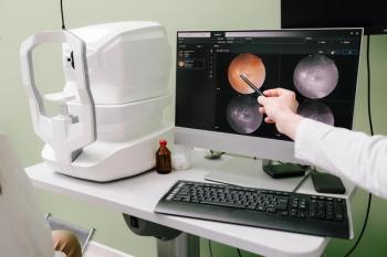
Chous: So what I use in my practice is a handheld electroretinography device that allows you, in about 3 minutes or so, to capture electrical signals from the retina when it's exposed to different levels of stimulus. It also measures the pupil response to the same stimulus. And recently, this just got published about a month ago, this technology was compared for predicting which patients with non-proliferative diabetic retinopathy were going to progress on to sight-threatening disease over the next year. And what was found is that this particular global index of retinopathy severity, ascertained by this objective measure of retinal function, was the most predictive single measure for who was going to need treatment in the next year. It beat out optical coherence tomography and geography; in fact, don't tell Dr Rodman. I said that it was better; it was better than an ultrawide field, fluorescein angiography, and all 3 of those that I just alluded to were far superior to grading color fundus photograph by retina specialist. So this comes from a study called TIME-2b, which used subcutaneous medication for preventing progression of diabetic retinopathy. The drug failed, so it didn't meet its primary end point, but they did look at different technologies for assessing who was going to get worse, and this particular electrophysiological measure called the DR Score, Diabetic Retinopathy Score, was the winner hands down compared to the other technologies. I think that's really exciting for us in primary primary eye care, because it allows us to decide which patients need to be followed more closely, and perhaps which patients have worse the disease than we would have thought otherwise, so that we can maybe get them in the hands of a retina specialist, if that's what they need.
So in what you're seeing now, which patients are the ones that are needing more close monitoring or more care?
Chous: Well, certainly based on classic criteria for grading patients that have more than moderate, non-proliferative retinopathy or any degree of diabetic macular edema, need to be followed much more closely. Now, what this technology allows me to do is to say, rather than just off the cuff saying, "Well, this patient I want to see back in a year or 6 months, because I see some non-proliferative changes, this patient has really highly abnormal retinal function, and it's based on this global score." So those are patients that I'm going to be seeing back sooner, and I'm going to give a couple of case examples of that in both of the talk that I presented this year at the Academy.
Was there anything else about that particular session that you wanted to highlight?
Chous: Just that Dr Rodman showed some awesome cases of using OCTA to really show us that patients who have sight-threatening retinopathy right now are sometimes missed when we rely on just simple fundus exam or standard retinal imaging techniques. So she's got some just wonderful cases demonstrating non-perfusion of the retina, as well as neovascularization that was not apparent on clinical exams. So the OCT really helped make the diagnosis. What I'm an advocate for, and I think Julie and I both advocate for, is using more advanced technologies in tandem with our normal clinical examination techniques to stratify patient risk better than we've been doing here before.
Newsletter
Want more insights like this? Subscribe to Optometry Times and get clinical pearls and practice tips delivered straight to your inbox.


