Clinical challenges in anterior segment ocular disease
Anterior segment ocular disease is one area where optometrists can really help their patients in need. Optometrists can treat anterior segment disease with topical medications and oral medications in many states. As primary eyecare providers, we are uniquely situated to care for our patients with anterior segment ocular disease. This is the third in a series of articles summarizing the Diplomate preparatory course by Primary Care Section of the American Academy of Optometry.
Dry eye
One common problem that presents to practically every optometrist’s office is dry eye (see Figure 1). Several years ago, a distinguished group of optometrists and ophthalmologists convened to arrive at a commonality of dry eye diagnosis and treatment.1 The international task force (ITF) coined the term “dysfunctional tear syndrome” to what we refer to as dry eye. This group actually preceded the Dry Eye Workshop (DEWS) and, while the term “dysfunctional tear syndrome” never caught on, many of the same recommendations were present in the DEWS report. Its guidelines broke the condition into 4 levels of severity and recommended treatment options for each:
• Level I may present with mild to moderate symptoms, and there may be mild to moderate conjunctival signs, but it is also possible there may be no signs.
• Level II patients may show moderate to severe symptoms, tear film signs, mild corneal punctate staining, and conjunctival staining.
• Level III symptoms will be severe, including marked corneal punctate staining, central corneal staining, and filamentary keratitis.
• Level IV patients experience extremely severe symptoms, possibly to the point of needing to alter their lifestyles. Look for severe corneal staining, erosions, and conjunctival scarring in this set of patients.
The treatment algorithm development of the ITF panel begins with patient education of her condition, changes in her environment, and attention to her systemic medications as it relates to her dry eye. Preserved artificial tears and allergy control is the first-line recommendation from mild or Level I dysfunctional tear syndrome. Nutritional supplements, cyclosporine A (Restasis, Allergan), and secretagogues are added at level II; oral tetracyclines and punctal occlusion at Level III; and systemic anti-inflammatories and acetylcysteine (Mucomyst, Bristol-Myers Squibb) at Level IV. While this is a very regimented protocol, obviously we cannot treat each patient in such a cookbook manner. It is incumbent upon each of us to treat every patient on an individual basis.
Hyperosmolarity has been described in the Tear Film and Ocular Surface Society (TFOS) dry eye report as a primary marker of tear film integrity and the key component of dry eye disease. 2 When the quantity or quality of secreted tears is compromised, increased rates of evaporation lead to a more concentrated tear film measured as increased tear osmolarity, which places stress on the corneal epithelium and conjunctiva. With filamentary keratitis, consider the use of acetylcysteine. This mucolytic goes in the nebulizer to break up phlegm in asthmatics. It makes a nice drop to break up the filaments in filamentary keratitis when you mix it one-to-one with artificial tears. This needs to be prepared by your local compounding pharmacist. The one drawback is it has a strong odor.
Meibomian gland dysfunction (MGD)
Anterior blepharitis is an inflammatory condition of the outer portion of the eyelids and is often secondary to infection or associated with acne rosacea or seborrheic dermatitis. Posterior blepharitis is an inflammation of the inner portion of the eyelids and is associated with altered composition of the meibomian gland secretions.3
Signs and symptoms of posterior blepharitis include the characteristic plugged meibomian gland openings and the foamy or soapy tear film that is pathognomonic for the disease. The normal meibomian gland secretions change in essence from an unsaturated fat to a saturated fat.
The normal monoglycerides and diglycerides are more solid in meibomian gland dysfunction, which leads to orifice obstruction and plugging of the glands. The white cheesy plugs and toothpaste-like exudate are degraded triglycerides. The monoglycerides and diglycerides are pro-inflammatory, which leads to the inflammation associated with meibomian gland disease.
We are all familiar with the traditional treatments for blepharitis: the warm compresses, lid scrubs and topical antibiotic therapies. Newer therapies include the omega-3 antioxidants and oral tetracycline class drugs, used more for anti-inflammatory activity than their antibiotic effect. Traditional treatments are now augmented by the use of azithromycin 1% topical drop (AzaSite, Akorn), massaged into the eyelids twice a day for 2 days, then once a day for 28 days.
GPC/VKC
Giant papillary conjunctivitis (GPC) is not a true allergic reaction, as is the case with seasonal allergic conjunctivitis (SAC), atopic keratoconjunctivitis (AKC), and vernal keratoconjunctivitis (VKC). GPC is caused by the repeated mechanical irritation of the papillary conjunctiva, and it is aggravated by concomitant allergy.
We're all familiar with the clinical presentation of GPC: mild lid hyperemia, thick mucus buildup, and uniform flat papillae on the superior tarsal plate. Treatment includes removing the offending cause, topical histamines/mast cell stabilizers, and topical steroids. The combination mast cell/antihistamine drops, while recommended for once-daily dosing, are quite safe and can be used more often as needed.
VKC is a chronic inflammation occurring most frequently during the spring and summer months, due to a normal seasonal increase in allergens in the air. It can also be caused by an allergic reaction to other irritants, such as chlorine in swimming pools, cigarette smoke, and ingredients in cosmetics. It is a disease predominately affecting young boys, and research has shown these patients may have a histaminase deficiency.4
The patient complains of intense itching and tearing and may complain of a hot feeling in his eyes, along with photophobia. Manage this presentation with an oral antihistamine, a combination antihistamine/mast cell stabilizer several times a day, and topical steroids. A cold pack will help relieve swelling and redness and will make the patient feel better.
If there is a corneal epithelial defect, a bandage contact lens can be employed. If a bandage contact lens is used, cover with a topical antibiotic. These patients can resolve within a few weeks of maximum medical therapy, but they're looking at a maintenance medication like a combination mast cell stabilizer/antihistamine drop.
Microbial keratitis
What microbe is commonly found in corneal ulcers (see Figure 3)? The Steroids for Corneal Ulcers Trial (SCUT) study provides interesting data. The SCUT study was the first large, prospective randomized clinical trial assessing the impact of adjunctive topical corticosteroids in patients with bacterial corneal ulcers.5 The most commonly isolated Gram-positive microbe in the SCUT study was Strep pneumoniae, and the most common Gram-negative microbe was Pseudomonas aeruginosa. Contact lens wear is a common risk factor for corneal ulcers in United States, in contrast to agricultural work being the most common risk factor in India, where the study was conducted.
What about the use of corticosteroids in corneal ulcers? We've all seen these used, but is that good medicine? The results of the SCUT study can guide us. The SCUT study found no significant difference in 3-month best spectacle corrected visual acuity (BCVA) between patients receiving topical corticosteroid or placebo as adjunctive therapy in the treatment of bacterial corneal ulcers. The results of the SCUT study demonstrate no obvious benefit in using corticosteroids in the overall study population.
An intriguing finding of the SCUT study was that subgroup analysis demonstrated a benefit in 3-month BCVA using corticosteroids in corneal ulcers with the greatest severity at presentation.
Corticosteroid treatment was associated with the benefit in visual acuity (VA) in the subgroups with the worst VA and central corneal ulcer location at baseline. These subgroup analyses suggest that patients with severe ulcers, those who have the most to gain in terms of VA, may benefit from the use of corticosteroids as adjunctive therapy. It's ironic that these central ulcers are the ones we are the most afraid to use steroids on.
Fuch’s dystrophy
Fuch's dystrophy is an autosomal dominant inherited disease that affects women greater than men.6 It typically presents in the fifth to sixth decade of life as multiple central corneal guttata (excrescences of Descemet's membrane) associated with pigment dusting on the endothelium. The condition spreads from the center toward the periphery.
As the endothelial cells fail, the remaining cells enlarge to cover the gap. With the reduced number of endothelial cells, the endothelial pump function suffers.
This leads to corneal edema and loss of VA. Vision is typically worse upon awakening because of the swelling induced by nighttime lid closure. In more advanced stages, the epithelial microcysts later coalesce forming bullae, which can rupture, causing foreign body sensation and pain, as well as exposing the cornea to the danger of infectious keratitis.
Treatment for Fuch’s include sodium chloride 5% eye drops instilled 4 to 6 times during the day, especially in the early hours of the day and less frequently in the evening. Sodium chloride ointment (Muro 128, Bausch & Lomb) is used at bedtime.
Using a hairdryer, kept at arm's distance, can be used to blow warm air over the cornea for 5 to 10 minutes upon awakening. Dehydrating the cornea in this manner may improve the vision of the patient for some time. Lowering the intraocular pressure is useful when it is even mildly elevated. It occasionally helps even when the pressure is normal, especially in borderline cases of corneal decompensation. Topical carbonic anhydrase inhibitors should be avoided because they hinder the activity of the endothelial pump.
Failing vision in the presence of epithelial edema and stromal haze, which does not resolve with treatment, is an indication for surgery. There are 3 surgical options for the Fuch’s patient: penetrating keratoplasty (PK) has long been the standard treatment a future the field dystrophy.
In the last few years, major advances have made replacement of the endothelial layer possible without disturbing normal anterior structures of the cornea using endothelial keratoplasty. Descemet's stripping endothelial keratoplasty (DSEK) involves the transplant of healthy endothelial layer along with minimal posterior corneal stroma.7 Descemet's membrane endothelial keratoplasty (DMEK) is the transplant of endothelial cells along with Descemet's membrane only.
Patients who undergo DSEK regain early and more superior VA than patients who undergo PK due to lack of surface sutures. These eyes are structurally stronger and more resistant to postoperative dramatic injury, and no suture related graft infection or graft rejection occurs.
Floppy eyelid syndrome
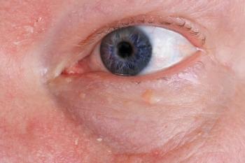
Floppy eyelid syndrome typically develops in obese patients with extremely lax eyelids, which evert spontaneously or with minimal manipulation (see Figure 4).
Classically, the eyelids evert and rub on the pillow or sheets while the patient is asleep, which is the cause of the papillary conjunctivitis and exposure keratopathy exhibited when the patient presents complaining of foreign body sensation and ocular irritation. Patients with this disorder need a referral to an internist or pulmonologist for evaluation of sleep apnea syndrome, which can be associated with floppy eyelid syndrome. Obstructive sleep apnea is a potentially fatal disorder.
Frequent episodes of apnea can lead to systemic and pulmonary hypertension and ultimately congestive cardiomyopathy. Obstructive sleep apnea is associated with other serious ocular disorders, such as keratoconus from rubbing the eyes, glaucoma, ischemic optic neuropathy, and papilledema secondary to increased intracranial pressure.8 Floppy eyelid syndrome is often unrecognized, and you can do your patients a real service by recognizing this condition and making the appropriate referral.
Opportunity to excel
Anterior segment ocular disease is one area in our practice where we can really excel. Our patients present to our office for us to care for these conditions regularly, and proper treatment is a great practice builder as a healthy, happy patient can generate tremendous word of mouth and loyalty.ODT
References
1. Behrens A, Doyle JJ, Stern L, Chuck RS, et al. Dysfunctional tear syndrome: a Delphi approach to treatment recommendations. Cornea. 2006 Sep;25(8): 900–7.
2. Dry Eye Workshop Report. The Ocular Surface. 2007 Apr;5(2).
3. Nichols KK. Blepharitis and dry eye: a common, yet complicated combination. Rev Optom. August 17, 2010.
4. Ono SB, Abelson MB. Clin Therapeutics 2004; 26 (8): 118-122.
5. Srinivasan M, Mascarenhas J, Rajaraman R, Ravindran M, et al. Corticosteroids for bacterial keratitis: the Steroids for Corneal Ulcers Trial (SCUT). Arch Ophthalmol. 2012 Feb;130(2): 143-50.
6. Lang GK, Naumann GO. The frequency of corneal dystrophies requiring keratoplasty in Europe and the USA. Cornea. 1987;6(3): 209-11.
7. McVeigh K, Somish KS, Reddy AR, Vakros G. Retained Descemet’s membrane following penetrating keratoplasty for Fuch’s endothelial dystrophy: a case report of a post-operative complication. Clin Ophthalmol. 2013;7:1511-4.
8. McNab AA. The eye and sleep apnea. Sleep Med Rev. 2007 Aug;11(4): 269-76.
Newsletter
Want more insights like this? Subscribe to Optometry Times and get clinical pearls and practice tips delivered straight to your inbox.


