

|Articles|March 28, 2023
- March digital edition 2023
- Volume 15
- Issue 03
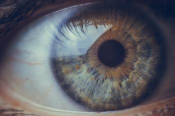
Diagnosing and treating optic nerve head drusen
Technologies that improve treatment of ONHD and similar pathologies.
Advertisement
Optic nerve head drusen (ONHD) are acellular hyaline deposits that aggregate within the prelaminar tissue of the optic nerve.1 Usually diagnosed early in life, ONHD are most likely to change in childhood but usually remain stable through adulthood.2 ONHD are estimated to occur in about 2.4% of patients.3 Many patients with ONHD are asymptomatic and their vision continues unaffected; however, ONHD can cause vision loss, nonarteritic anterior ischemic optic neuropathy (NAION), and peripapillary choroidal neovascular membranes. Although ONHD are usually an incidental finding during a routine exam, up to 10% of patients with them report transient visual obscurations.4 When located in more superficial layers of the optic nerve, drusen tend to take a pale color and can appear small as well as large and lobulated. When located deeper within the optic nerve, they can cause the optic nerve to take on a swollen or elevated appearance.1 Superficially located ONHD may be diagnosed with fundoscopy, but those more deeply located require additional testing to rule out other ocular pathology.
ONHD may be difficult to detect and properly diagnose on fundoscopic examination alone, as the optic nerve may appear elevated and the disc margins blurred, mimicking the appearance of papilledema. Differentiating optic disc drusen from papilledema, optic nerve edema, and other causes of pseudopapilledema is essential because each diagnosis entails different levels of severity and management protocols.
Imaging ONHD
Although enhanced depth imaging optical coherence tomography (EDI-OCT) used to be the gold standard for ONHD detection, B-scan ultrasonography is invaluable for its evaluation because it reveals elevated and highly reflective calcified drusen at the optic nerve head.2 This is an important tool, especially if the drusen are buried, because it allows visualization of their posterior borders. Nevertheless, ultrasonography in the context of disc drusen has several limitations. B scans don’t provide detailed information about the integrity of the optic nerve and surrounding tissue. Given that drusen tend to calcify at around age 12,5 they have yet to do so in many pediatric patients and are often buried, making detection via B scan difficult, if not impossible.
To differentiate optic nerve head drusen from papilledema, lower the gain setting and assess signal intensity.5 In the presence of ONHD, deposits remain hyperreflective, whereas with papilledema, the signal intensity decreases along with the gain setting. Ultrasonography may reveal a widening of the optic nerve sheath because of increased fluid within the sheath and the classic crescent shadow.
OCT also allows clinicians to confidently rule out disc edema. Patients with disc edema will have a thickened RNFL and surrounding subretinal fluid.1 OCT scans of the optic nerve head should be performed annually in patients known to have ONHD to monitor changes and potential RNFL damage.
Another effective tool for diagnosing and monitoring ONHD is fundus autofluorescence (FAF), which utilizes the blue excitation filter and green imaging filter used in fluorescein angiography.6 These filters make optic nerve head drusen appear bright and lobulated, allowing for easy viewing.1
ONHD-associated NAION
Recent studies have expanded our understanding of the complications associated with ONHD. One prospective study compared 13 patients with optic disc drusen associated nonarteritic anterior ischemic optic neuropathy (ODD-AION) and 14 with nonarteritic anterior ischemic optic neuropathy (NA-AION) and found that those with ODD-AION were younger and had fewer vascular risk factors.1 According to the researchers, “findings suggest that [ONHD] is an independent risk factor for developing anterior ischemic optic neuropathy.”3
Another study compared the optic nerve head anatomy of patients with ODD-AION and NA-AIONand found that 82% of those with ODD-AION had deep rather than superficial ONHD. They concluded that anatomical location, not size, was important for determining a patient’s risk of developing ODD-NAION. The study echoed existing literature, suggesting that ONHD is an independent risk factor for NA-AION and noted that ODD-AION patients “are younger at the time of diagnosis and have fewer vascular risk factors” than NAION patients.7
These studies have significant clinical implications and demonstrate the importance of proper diagnosis.
Managing ONHD
As noted above, atypical progression and associated complications of ONHD cannot be ruled out. Thus, once a proper diagnosis has been made, patients with ONHD must be monitored closely to assess RNFL damage and related visual field defects, peripapillary neovascular membranes, and ODD-AION.7
There is currently no treatment for patients with visual field loss secondary to ONHD.2 Some retrospective studies recommend the use of medications that lower intraocular pressure; however, there are no controlled clinical studies to support this approach, and a consensus among clinicians has yet to be reached. Patients with ONHD should be dilated yearly for fundus evaluation and autofluorescence, OCT scans, and visual field exams. It is important to note that many of these patients have varying degrees of visual field defects.2 About 50% of children and 80% of adults with ONHD have an underlying visual field defect, typically enlarged blind spots.8 And although patients are generally asymptomatic for these defects, they require close monitoring. Patients rarely experience profound vision loss, but the ones who do usually have it secondary to NAION.8 As previously mentioned, patients with ONHD will often demonstrate autofluorescence in the areas within the optic nerve where the drusen are present.
Technological advances have been critical to the unearthing of ONHD, with recent studies suggesting ONHD as an independent risk factor for NA-AION in younger patients with fewer vascular conditions than others with NA-AION. In 2019, the Center for Optic Disc Drusen was created at Stanford School of Medicine to further propel our understanding of ONHD and its implication in blinding eye diseases.9 The future is bright for the use of technology improving outcomes for these patients.
References
1. Palmer E, Gale J, Crowston JG, Wells AP. Optic nerve head drusen: an update. Neuro-Ophthalmology. 2018;42(6):367-384. doi:10.1080/01658107.2018.1444060
2. American Academy of Ophthalmology. Optic nerve head drusen. EyeWiki. September 11, 2022. Accessed November 20, 2022. https://eyewiki.aao.org/Optic_Nerve_Head_Drusen
3. Malmqvist L, Bursztyn L, Costello F, et al. The Optic Disc Drusen Studies Consortium recommendations for diagnosis of optic disc drusen using optical coherence tomography. J Neuroophthalmol. 2018;38(3):299-307. doi:10.1097/WNO.0000000000000585
4. Ahmed H, Khazaeni L. Optic disc drusen. StatPearls. Updated May 14, 2022. https://www.ncbi.nlm.nih.gov/books/NBK580547/
5. Chang MY, Pineles SL. Optic disk drusen in children. Surv Ophthalmol. 2016;61(6):745-758. doi:10.1016/j.survophthal.2016.03.007
6. Fraser JA, Bursztyn LL. Optical coherence tomography in optic disc drusen. Ann Eye Sci. 2020;5:5. doi:10.21037/aes.2019.12.03
7. Johannesen RG, Lykkebirk L, Jørgensen M, Malmqvist L, Hamann S. Optic nerve head anatomy and vascular risk factors in patients with optic disc drusen associated anterior ischemic optic neuropathy. Am J Ophthalmol. 2022;242:156-164. doi:10.1016/j.ajo.2022.06.016
8. Lee KM, Woo SJ, Hwang JM. Factors associated with visual field defects of optic disc drusen. PLoS ONE. 2018;13(4):e0196001. doi:10.1371/journal.pone.0196001
9. Introducing: the new center for optic disc drusen at Stanford. Stanford Medicine. Accessed November 19, 2022. https://med.stanford.edu/optic-disc-drusen/meetings-and-lectures/2020/ODD.html
Articles in this issue
about 3 years ago
New topical therapies for dry eye diseaseabout 3 years ago
Educating patients on refractive surgery optionsover 3 years ago
My practice journeyover 3 years ago
Artificial tear ingredients that mimic the real thingover 3 years ago
Supporting glaucoma patients beyond the exam roomover 3 years ago
Management of chronic allergic eye diseaseover 3 years ago
Breaking into lens techAdvertisement
Related Content
Advertisement


Latest CME


Advertisement
Advertisement
Trending on Optometry Times - Clinical News & Expert Optometrist Insights
1
Epion Therapeutics completes phase 3 trials of EpiSmart epithelium-on cross-linking for keratoconus
2
Euclid Vision receives FDA clearance for Be Free Day toric contact lenses
3
Sparkling retina: Multimodal imaging in Bietti crystalline dystrophy
4
Oculis enrolls first patient in licaminlimab PREDICT-1 DED trial
5