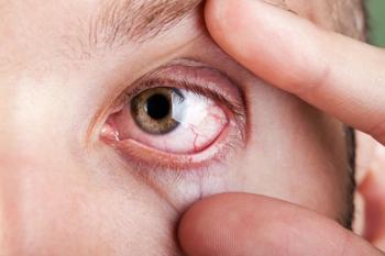
Evolution of diagnosis methods will preserve vision, prevent AMD progression
Traditional methods of diagnosing AMD are not effective, now that there are been significant advances in treatment. Early diagnosis is imperative in order for patients to have better outcomes, including preserving vision and preventing disease progression, from dry to wet AMD.Diagnostics are evolving to diagnosis AMD earlier.
Seattle-Traditional methods of diagnosing age-related macular degeneration (AMD) are not effective, now that there are been significant advances in treatment. Early diagnosis is imperative in order for patients to have better outcomes, including preserving vision and preventing disease progression, from dry to wet AMD.
In a presentation given at the annual meeting of the American Academy of Optometry, Steven Ferrucci, OD, pointed out that since 2004, when pegaptanib sodium injection (Macugen, EyeTech/Pfizer) was approved for the treatment of neovascular AMD, there are now better options for therapy. “We need to talk about new and better ways of diagnosis and getting AMD diagnosed earlier.”
Diagnostics are evolving as well, making this a reality, and optometrists have an important role to play in this evolution, said Dr. Ferrucci, an associate professor and chief of optometry, Sepulveda VA Medical Center, Sepulveda, CA.
One tool is the AdaptDx (MacuLogix), which can monitor “dark adaptation,” or the transition from day vision to night vision. Dark adaptation impairment is the clinical hallmark of early AMD.
“If you dark adapt in less than 6.5 minutes, you are probably normal,” he explained, noting that one recent study found that dark adaptation correctly identified 90.6% of AMD cases.
Advantages of the AdaptDx are no pre-adaptation required; it is easy to operate; and doesn’t place a burden on the patient. (The device is not currently cleared for sale as a diagnostic tool for AMD by the FDA.)
Fundus autofluorescence imaging (FAF) is another noninvasive diagnostic technique, and can be used for the deposition of lipofuscin in the retinal pigment epithelium (RPE). Lipofuscin deposition normally increases with age, but may also occur from RPE cell dysfunction or an abnormal metabolic load on the RPE.
“Variation seen in FAF may precede retinal changes and may be prognostic for those patients who will continue to develop vision loss,” said Dr. Ferrucci.
It has now been established that AMD has definite genetic components, and known markers account for about 70% of the population as a risk. The other 30% is environment/lifestyle.
The two most important genes are CFH and ARMs 2/HTRA1, and testing is now available for these genes, he explained. “So if you look at the patient’s genetic make-up and then at the risk factors, the risk can be better estimated,” Dr. Ferrucci said.
Knowledge of genetic risk is important as patients can then be counseled about what can be done to reduce that risk, he added.
Newsletter
Want more insights like this? Subscribe to Optometry Times and get clinical pearls and practice tips delivered straight to your inbox.


