Extended ophthalmoscopy is a must for diagnosing, treating vitreous floaters
Vitreous floaters are a common patient complaint, but because floaters are typically benign and the treatment options are limited, most can be managed with proper patient education and reassurance.
"The important thing that optometrists can do is to be knowledgeable and competent doing the history and make sure to do a good differential diagnosis. Don't be too dismissive of floaters," Dr. Schoen said. "This could be a floater that the patient has had for 2 decades the majority of time, but your history should tease that out."
The nature of floaters
Posterior vitreous detachment symptoms may include new or increasing floaters as well as photopsia. Occurrence in one eye foreshadows increasing probability of occurrence in the fellow eye. Myopia is associated with peripheral vitreous detachment as well, Dr. Schoen explained.
"For example, patients with lid ptosis, dry eye, or collarets may report particles in their vision. It's part of our job in taking a good history to tease those out," he said.
Doing the work-up
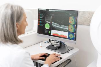
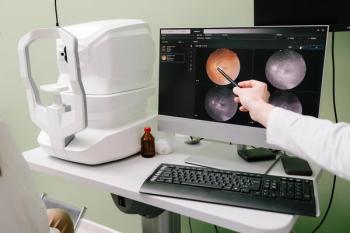
"Don't look through the vitreous; look at the vitreous," Dr. Schoen said. So he advised against initially relying upon untrasound, optical coherence tomography, and other newer diagnostic technologies. Dilated fundus examination extending out to the equator of the globe and beyond is the gold standard, he said. Pay attention to the vitreous base as a potential location for hemorrhage and utilize scleral depression to do surveillance for occult flaps and tears of the peripheral retina.
"The biggest lapse clinicians are guilty of is failing to do a meticulous evaluation of the peripheral retina through a dilated pupil. Just dilate the patient and look, and do so very carefully," he said. "If the floaters are so thick that it's difficult to visualize the posterior pole, consider ordering an ultrasound. This will offer you an image and give you an idea of what is going on."
He noted most experts advise doing dilated extended ophthalmoscopy as soon as possible, particularly when a patient has persistent photopsia, clouded vision, or sudden onset of either one or two large floaters or myriad smaller floaters.
Newsletter
Want more insights like this? Subscribe to Optometry Times and get clinical pearls and practice tips delivered straight to your inbox.


