

|Articles|July 7, 2023
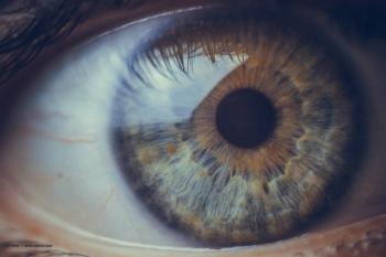
Fixational eye movements: The eye motion no one is talking about, but should be
Author(s)Jacqueline Theis, OD, FAAO
These 3 types of fixational eye movements play a vital role in our visual perception, cognitive function, and are instrumental for our ability to see the world around us.
Advertisement
We spend over 80% of our visual day fixating our gaze on stationary objects, but when was the last time you thought about or evaluated your patient’s fixational stability? Fixation is not a static passive state, but rather an active neural process.
These microscopic flickers of motion may seem insignificant, as they are unnoticed by most people and undetectable on standard clinical exam; however, these minute eye movements play a vital role in our visual perception, cognitive function, and are instrumental for our ability to see the world around us with 20/15 clarity and precision.
As foveate animals, humans necessitate fixational eye movements because of the uneven distribution of photoreceptors in the retina. Our eyes need to be precisely pointed at objects of visual interest so that the light image from these objects is focused on the fovea. If the light is focused parafoveally or peripherally, our visual acuity begins to decline. Thus, fixational eye movements help maintain eye alignment.
An additional function of fixational eye movements is to prevent retinal neural adaptation, a phenomenon where the sensitivity of our retinal receptors decreases over time when exposed to a constant stimulus. Thus, the eyes need to constantly microscopically move or our visual system will desensitize to the visual stimulus. Quite the conundrum, because if the eyes move too much during fixation, then the patient will lose clarity and complain of blur and/or oscillopsia, but if they don’t move enough the patient will complain of visual fading.1,2
There are 3 types of fixational eye movements: microsaccades, tremor, and drift. Microsaccades are fast, jerk-like eye movements that occur about 1-4 times a second. They are the largest of fixational eye movements, moving the image across several dozen to hundreds of photoreceptors. Drift, on the other hand, refers to the slow, smooth motion that occurs in between microsaccades, moving the image across about a dozen photoreceptors. Lastly, tremor, the smallest fixational eye movement, refers to the high frequency vibration of the eyes that occurs simultaneously with drift, and moves the image over about the diameter of a foveal cone.3-5
Technological advancement in the last few decades has led to a revival of interest in fixational eye movements and it is one of the fastest moving fields in visual neuroscience today. Anatomically, fixational eye movements require a vast portion of brain circuitry including every lobe of the brain, brainstem, cerebellum, thalamus, basal ganglia, cranial nerves, and afferent visual pathway.6 Thus, fixational eye movements are susceptible to ophthalmologic and neurologic insults.
Researchers have found abnormalities in fixational eye movements in mild traumatic brain injury,7 multiple sclerosis,8 mild cognitive impairment, Alzheimer’s disease,9 Parkinson’s disease,10 amblyopia,11 maculopathy,12 and schizophrenia.13 These findings suggest that analyzing fixational eye movements could potentially serve as a non-invasive tool for early detection and monitoring of certain neurological disorders.
References
- Troxler, D. (1804). Ueber das Verschwinden gegebener Gegenstande innerhalb unseres Gesichtskreises. In: Himley, K. and Schmidt, J.A. (Eds.), Ophthalmologische Bibliothek, Springer, Jena, pp. 1–53.
- Martinez-Conde S, Macknik SL, Troncoso XG, Dyar TA. Microsaccades counteract visual fading during fixation. Neuron. 2006;49(2):297-305. doi:10.1016/j.neuron.2005.11.033
- Leigh JR, DS Zee. The Neurology of Eye Movements, 5th ed, Contemporary Neurology Series (New York, 2015; online edn, Oxford Press.
- Martinez-Conde S. Fixational eye movements in normal and pathological vision. Prog Brain Res. 2006;154:151-176. doi:10.1016/S0079-6123(06)54008-7
- Wong AMD. Eye Movement Disorders. Oxford Press. 2008.
- Coiner B, Pan H, Bennett ML, et al. Functional neuroanatomy of the human eye movement network: a review and atlas. Brain Struct Funct. 2019;224(8):2603-2617. doi:10.1007/s00429-019-01932-7
- Leonard BT, Kontos AP, Marchetti GF, et al. Fixational eye movements following concussion. J Vis. 2021;21(13):11. doi:10.1167/jov.21.13.11
- Sheehy CK, Bensinger ES, Romeo A, et al. Fixational microsaccades: A quantitative and objective measure of disability in multiple sclerosis. Mult Scler. 2020;26(3):343-353. doi:10.1177/1352458519894712
- Kapoula Z, Yang Q, Otero-Millan J, et al. Distinctive features of microsaccades in Alzheimer's disease and in mild cognitive impairment. Age (Dordr). 2014;36(2):535-543. doi:10.1007/s11357-013-9582-3
- Otero-Millan J, Schneider R, Leigh RJ, Macknik SL, Martinez-Conde S. Saccades during attempted fixation in parkinsonian disorders and recessive ataxia: from microsaccades to square-wave jerks. PLoS One. 2013;8(3):e58535. doi:10.1371/journal.pone.0058535
- Kang SL, Beylergil SB, Otero-Millan J, Shaikh AG, Ghasia FF. Fixational Eye Movement Waveforms in Amblyopia: Characteristics of Fast and Slow Eye Movements. J Eye Mov Res. 2019;12(6):10.16910/jemr.12.6.9. Published 2019 Jul 5. doi:10.16910/jemr.12.6.9
- Kumar G, Chung ST. Characteristics of fixational eye movements in people with macular disease. Invest Ophthalmol Vis Sci. 2014;55(8):5125-5133. Published 2014 Jul 29. doi:10.1167/iovs.14-14608
- Egaña JI, Devia C, Mayol R, et al. Small Saccades and Image Complexity during Free Viewing of Natural Images in Schizophrenia. Front Psychiatry. 2013;4:37. Published 2013 May 20. doi:10.3389/fpsyt.2013.00037
Advertisement
Related Content
Advertisement


Latest CME


Advertisement
Advertisement
Trending on Optometry Times - Clinical News & Expert Optometrist Insights
1
Epion Therapeutics completes phase 3 trials of EpiSmart epithelium-on cross-linking for keratoconus
2
Euclid Vision receives FDA clearance for Be Free Day toric contact lenses
3
Sparkling retina: Multimodal imaging in Bietti crystalline dystrophy
4
Oculis enrolls first patient in licaminlimab PREDICT-1 DED trial
5