IKA 2024: The urgency in keratoconus management
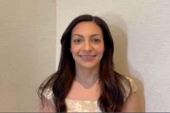
Kourtney Houser, MD, and Kendall Donaldson, MD, MS, give an overview of the important role topography and tomography play in catching keratoconus early in their patients.
In their talk, "Urgency in Keratoconus Management: Early Detection Can Prevent Vision Loss," Kourtney Houser, MD, and Kendall Donaldson, MD, MS, review the methods to catching keratoconus early, including tomography and topography to properly assess the changes in the anterior cornea. They gave this talk at the International Keratoconus Academy Keratoconus Symposium.
Video Transcript
Editor’s note - The following transcript has been lightly edited for clarity.
Kourtney Houser, MD:
Hi, I'm Kourtney Houser at Duke University in North Carolina.
Kendall Donaldson, MD, MS:
And I'm Kendall Donaldson from Miami, Florida at the Bascom Palmer Eye Institute. Today, Kourtney and I did a presentation together on early diagnosis of keratoconus. And we talked about the prevalence of keratoconus, which at 1 time was thought to be about 1 in 2000. But now we know better; it's actually closer to 1 in 250 or 1 and 300 patients. So over the last decade, we've learned a lot about how prevalent keratoconus truly is. And that early diagnosis is just so important. One of the main things we use for the diagnosis is topography.
Houser:
We also reviewed topography, tomography, some of the older indices that sort of started this wave of diagnosis early and then some of the key indicators in tomography that might help us diagnose keratoconus before we lose vision when the anterior cornea changes. And all this is so important because we now have corneal collagen cross-linking, where if we catch patients early, we could stabilize them before they lose spectacle corrected visual acuity. We also reviewed biomechanics, which hopefully someday we'll have new tests that we could measure corneas to actually detect their innate corneal strength instead of just estimating patients that might be at a risk for ectasia. And this may be a wave of the future for us to be able to have even more diagnostic tools available to us.
Donaldson:
Our goal for the future of keratoconus diagnosis would be to always avoid scarring, and changes in the typography, and avoid the need for corneal transplantation. We talked about some of the nuances on corneal tomography and learning to read to corneal tomography better. We're very fortunate to have things like the Belin-Ambrosio display that helps us diagnose keratoconus a little bit earlier. So we have many different tools like corneal hysteresis, you know, ways to measure the cornea and just teaching everyone whether they're general practitioners, family practitioners, pediatric ophthalmologists, general ophthalmologists, and of course the corneal practitioner to all work together to help diagnose these patients earlier, is really key to help make the diagnosis and avoid some of the horrible sight quality that we see in our keratoconus patients.
Houser:
And in the spirit of such a collaborative meeting, like we have here, we think that it's really important for us to continue to collaborate, as optometrists see so many of these patients early on to help us continue to catch patients earlier in earlier so that we can treat with cross-linking at earlier ages to hopefully prevent spectacle corrected vision loss in the future.
Newsletter
Want more insights like this? Subscribe to Optometry Times and get clinical pearls and practice tips delivered straight to your inbox.


