
|Articles|January 1, 2010
Look into the future of glaucoma therapy
New surgical techniques, more effective intraocular pressure monitoring devices, and a better understanding of the value of neuroprotection will guide the management of glaucoma patients in the future.
Advertisement
Dr. McPherran co-presented with David Yang, OD, FAAO, at the annual meeting of the American Academy of Optometry.
"There are many exciting developments in the field of glaucoma," Dr. McPherran said. "We have new surgical management procedures that are working very nicely and that have the potential to replace older surgeries, such as trabeculectomies. There is interesting research looking into the aging process of the optic nerve, and the eventual emergence of implantable telemetry devices will enhance our ability to accurately take pressure readings from directly inside the eye."
Leading-edge technology
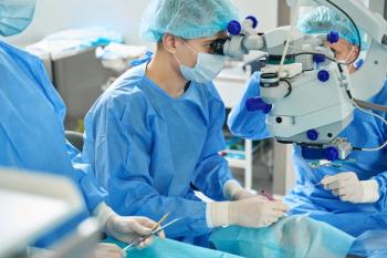
Dr. McPherran said he has co-managed a number of canaloplasty patients, and the 1- and 2-year IOP management results of those procedures have been good. Implantation of the new shunts may prove to be a valuable procedure for patients who cannot be compliant because of physical or mental disabilities, he said.
Preventing patients from needing surgical intervention, however, will remain a major treatment goal for optometrists. That will call for a high level of relative risk assessment from the profession, and will mean optometrists will need to stay up-to-date on new technologies that will become available to them.
"We're still using analog measurement instruments in a digital age," Dr. McPherran said. "There are many issues with how we take IOP measurements. Is the instrument we're using accurate? How do we measure nocturnal IOP? Also, the rigidity and thickness of the cornea changes over time because of age-related collagen cross-linking. So we really need to get inside the eye to know what's going on."
New devices, such as advanced compensating tonometers, are improving the ability to accurately and consistently measure IOP, but implantable devices hold the greatest promise, according to Dr. McPherran. Although implantable IOP monitoring devices will be valuable tools, the challenge remains how to power them. Realistically, they're still about 5 years away from joining the ranks of monitoring options, he said.
Neuro-optometry issues
Research into and a greater understanding of optic nerve damage and the role of neuroprotection also will increasingly guide treatment decisions.
"The cribiform plate appears to have a diaphragm or 'trampoline-type' effect," Dr. McPherran said. "It basically can move with some of the pressure variances within the eye. In younger eyes, the porosity of the plate is relatively softer, and the nerve fibers that go through those pores are not as easily damaged. But as the cribiform gets older, it becomes more cross-linked with glycation end products and becomes stiffer. It also continues collagen deposition over time that makes it thicker, and this thicker and stiffer plate may be part of what's causing the nerve damage."
Risk assessment
Cerebrospinal fluid pressure on the other side of the cribiform plate may play a role in ocular nerve damage, Dr. McPherran added. While this pressure can't be easily modified, awareness of this mechanism of action may prove to be a key when deciding which patients are at higher risk of optic nerve damage and are candidates for early treatment intervention.
Ultimately, it is this ability to conduct an accurate relative risk assessment that will keep glaucoma in the forefront of interesting challenges for optometrists.
"We're always looking for new techniques to help us refine our risk assessments and better determine which patients are at risk for optic nerve damage, and those who have already had damage. We know that damage is progressive, so early intervention is important," Dr. McPherran said.
Evaluation of Ocular Hypertension Treatment Study data on the number needed to treat reveals that optometrists and ophthalmologists currently treat or manage large numbers of ocular hypertensives, many of whom may not progress to glaucoma. Increasing use of techniques, such as imaging devices to monitor progression rates, will allow optometrists to arrive at better relative risk assessments, according to Dr. McPherran.
Cost-containment also will play a major role in the glaucoma therapy of the future.
"If you've kept patients well-managed, have diligently monitored their progression rates, and are holding progression down, then you may be able to continue with medical management. But the game is changing, and the name of the new game is going to be cost containment," Dr. McPherran said.
"It may turn that it's better to turn to surgical management with new procedures that may have a greater efficacy and financial benefit than the long-term use of medication. The traditional management of glaucoma patients may well change as we get our cost containment needs in place," he concluded.
Newsletter
Want more insights like this? Subscribe to Optometry Times and get clinical pearls and practice tips delivered straight to your inbox.
Advertisement
Related Content
Advertisement


Latest CME


Advertisement
Advertisement
Trending on Optometry Times - Clinical News & Expert Optometrist Insights
1
Early SPECTRUM results outline week 8 real-world outcomes with aflibercept 8 mg
2
The Vision Council report shows optical industry value growth amid lower volumes in 2025
3
Higher head elevation during sleep may be associated with higher IOP, study suggests
4
Myra Vision treats first patient in ADAPT trial for Calibreye TGT Surgical System
5