

|Articles|July 29, 2019
Measurements matter in cataract surgery, the new refractive procedure
Author(s)Jim Owen, OD, MBA, FAAO
Advertisement
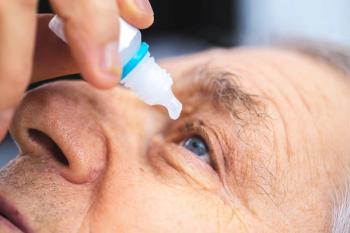
Prior to laser vision correction, ODs carefully examine the cornea. They evaluate the tear film, meibomian glands, curvature of both the front and back of the cornea; and measure overall corneal thickness.
Now that
Previously by Dr. Owen: How to determine an ideal LASIK candidate
New classification
At the start of my career,
Today,
Related: Choose your words carefully with surgery patients
Once a visually significant cataract has been identified in a motivated patient, the task of setting expectations begins. All clinical facts need to be gathered before the discussion of premium lens can start.
Findings as benign as posterior vitreous floaters can create visual disturbances that may impact the success of multifocal
The cataract patient should undergo a similar work-up of the ocular surface and its supporting structures as a contact lens patient or a laser vision correction patient. The significant difference is that positive findings do not prevent the procedure-they simply guide the
Related: Blog: Dive into the widening landscape of refractive surgery
Initial examination
Begin by evaluating structure before moving on to function.
I first image the meibomian glands using meibography. Showing patients meibography images and the absence of glands gets their attention to the importance of maintaining a healthy ocular surface.
Next, I evaluate the lid margin documenting erosion, telangiectasic vessels, and capped meibomian glands. By pressing on the meibomian glands, evaluate the quality of the meibum from absent, inspissated, or cloudy, to an olive oil consistency.
Related:
Noting how many glands produce meibum determines the productivity of those glands. There are treatments such as iLux (Alcon), LipiFlow (Johnson & Johnson Vision), and intense pulsed light (IPL), along with supportive therapy of hot compresses and omega-3 supplements.
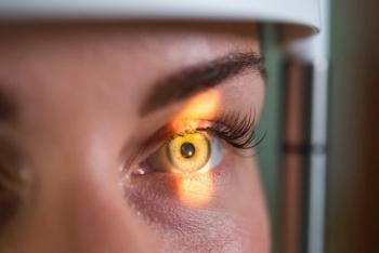
Fluorescein and lissamine green dyes provide information on both the structure and function of the tear film. Evaluate both cornea and conjunctival staining along with measuring tear break-up time (TBUT). An unstable tear film not only affects the quality of vision after the surgery but also the accuracy of the measurements that determine IOL power.
Study outcomes
In the Prospective Health Assessment of Cataract Patients’ Ocular Surface (PHACO) study, more than 50 percent of patients who were evaluated for cataract surgery had a TBUT of less than 5 seconds, and 75 percent had corneal staining.1
Hyperosmolar patients also had a greater variability in their keratometric measurements and a subsequent IOL power calculation of greater than 0.50 D difference.2 Therefore, the refractive “surprise” may be the simple result of a poor ocular surface.
Related:
As indicated, the corneal curvature plays a crucial role in the refractive power of the eye and determining IOL power. In fact, corneal astigmatism determines the power for a toric IOL. The corneal astigmatism is often similar to the refractive astigmatism-but not always.
In my experience, refractive spheres can have a significant amount of corneal astigmatism and- if not identified and addressed-can lead to postoperative refractive astigmatism.
The optical quality of the cornea impacts postoperative results. Therefore, corneal scars, epithelial basement dystrophy, and other corneal dystrophies need to be identified and have their potential impact on a patient’s vision discussed prior to surgery.
Additionally, keratoectasia can have a significant impact on the quality of vision after surgery. Pre-operative tomography can reveal forme fruste keratoconus (FFKC) and eyes that are not suited for corneal surgery to “fine-tune” a final refractive outcome. Those patients with even subtle corneal irregularities can experience poor postoperative quality of vision and less satisfaction, especially if they have a multifocal implant.3Related:
In a study of 19 patients with mild keratoconus, refractive astigmatism was significantly reduced with the implantation of a toric IOL and the spherical equivalent was within ±0.50 in 68 percent of the eyes.
Of note, the corneal astigmatism and total higher-order aberrations (HOA) were not significantly different.4 Therefore, a rigid
Conclusion
Today’s technology has transformed
Nevertheless, there are limitations that need to be identified and discussed prior to surgery. When properly evaluated, IOLs can provide patients with a quality of vision they have not experienced before, often without the aid of glasses or contact lenses. Those successful patients require evaluation and consultation from their referring doctors.
Read more refractive surgery content here
References:
1. Trattler WB, Majmudar PA, Donnenfeld ED, Stonecipher KG, Goldberg DF. The prospective health assessment of cataract patients’ ocular surface (PHACO) study: the effect of dry eye. Clin Ophthalmol. 2017 Aug 7;11:1423-1430.
2. Epitropoulos AT, Matossian C, Berdy GJ, Malhotra RP, Potvin R. Effect of tear osmolarity on repeatability of keratometry for cataract surgery planning. J Cataract Refract Surg. 2015 Aug;41(8):1672-7.
3. Montano M, Lopez-Dorantes KP, Ramirez-Miranda A, Graue-Hernandez EO, Navas A. Multifocal toric intraocular lens implantation for forme fruste and stable keratoconus. J Refract Surg. 2014 Apr;30(4):282-285.
4. Kamiya K, Shimizu K, Miyake T. Changes in astigmatism and corneal higher-order aberrations after phacoemulsification with toric intraocular implantation for mild keratoconus with cataract. Jpn J Ophth. 2016 Jul;60(4):302-8.
Newsletter
Want more insights like this? Subscribe to Optometry Times and get clinical pearls and practice tips delivered straight to your inbox.
Advertisement
Related Content
Advertisement


Latest CME


Advertisement
Advertisement