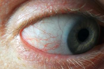
New research uncovers pathway for blink-assisting prostheses development
Specifically, the study found that the muscle that controls eyelid movement, or the orbicularis oculi, “contracts in complex patterns that vary by action and move the eyelid in more than just a simple up-and-down motion."
New discoveries regarding the muscle that controls blinking were recently uncovered by researchers at the University of California Los Angeles.1,2 The research, funded by the National Institutes of Health’s National Eye Institute (NEI) and published in the Proceedings of the National Academy of Sciences, could make way for developing blink-assisting prostheses in the future, according to a news release.
“The eyelid’s motion is both more complex and more precisely controlled by the nervous system than previously understood,” said study corresponding author Tyler Clites, PhD, an assistant professor of mechanical and aerospace engineering at the UCLA Samueli School of Engineering, in the release. “Different parts of the muscle activate in carefully timed sequences depending on what the eye is doing. This level of muscle control has never been recorded in the human eyelid. Now that we have this information in rich detail, we can move forward in designing neuroprostheses that help restore natural eyelid function.”
Specifically, the study found that the muscle that controls eyelid movement, or the orbicularis oculi, “contracts in complex patterns that vary by action and move the eyelid in more than just a simple up-and-down motion,” the release stated.1 The study involved 8 patients, all of whom had double eyelid morphology. Researchers evaluated how the muscle behaves during spontaneous blinks, protective rapid closure, and squeezed shut-eye motions, among other actions. Activity in the orbicularis oculi was recorded by inserting tiny wire electrodes into either the right or left eyelid. The procedure was completed by an ophthalmic surgeon. Researchers then used a motion-capture system to track eyelid movement in ultraslow motion, according to the release. Eyelid closure events were often around 100 ms in duration, with the capture rate of 400 frames per second providing approximately 40 samples for each trial.2
Ultimately, a set of 5 motion capture markers along the eyelid margin was tracked in 3 dimensions during 5 different eyelid behaviors, including spontaneous blink (an automatic, unconscious blink to keep the eye lubricated), voluntary blink (an intentional blink on command), reflexive blink (a rapid, involuntary blink triggered to protect the eye), soft closure (a slow eyelid descent, similar to the beginning of sleep), and a forced closure (deliberately squeezing the eyelids shut tightly). Researchers used 4 kinematic determinants to compare upper-eyelid kinematics between different eyelid behaviors, including onset medial traction as a percentage, reverberation as a percentage, eye closure as a percentage, and main sequence slope.2
“In this study, we sought to address these limitations by studying eyelid neuromechanics with high spatial and temporal resolution, via segmental intramuscular EMG and three-dimensional motion capture from markers along the eyelid margin,” the study authors, led by Jinyoung Kim, PhD, RN, FAAN, of the Department of Mechanical and Aerospace Engineering at the University of California Los Angeles, stated. “Our primary objective was to elucidate the differential activation patterns and resultant kinematics associated with each eyelid behavior, as a critical step toward mechanistically linking those neuromechanics to eyelid function. We also explored muscle excursion in the pretarsal orbicularis oculi, by tracking motion of the eyelid skin. These investigational tools have led us to findings about kinematic characteristics and temporospatial EMG activation patterns across different eyelid behaviors.”
“These results have significant implications for the development of neuroprosthetic systems to restore natural blink in persons with facial paresis,” the study authors stated.
References:
NEI-funded study reveals complex muscle control behind blinking and eyelid function. News release. National Eye Institute. August 11, 2025. Accessed August 18, 2025.
https://www.nei.nih.gov/about/news-and-events/news/nei-funded-study-reveals-complex-muscle-control-behind-blinking-and-eyelid-function Kim J, Shirriff A, Cornwell JN, et al. Human eyelid behavior is driven by segmental neural control of the orbicularis oculi. Proceedings Nat Academy Sci. 2025;122(32):e2508058122.
https://doi.org/10.1073/pnas.2508058122
Newsletter
Want more insights like this? Subscribe to Optometry Times and get clinical pearls and practice tips delivered straight to your inbox.


