
|Articles|July 11, 2020
- July digital edition 2020
- Volume 12
- Issue 07
Study finds FDT visual field studies are effective glaucoma detectors
Author(s)Benjamin P. Casella, OD, FAAO
Frequency doubling technology may become the new holy grail of glaucoma imaging.
Advertisement
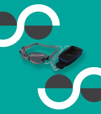
Frequency doubling technology (FDT) has been around for decades with visual field testing.1 The science centers around frequency.2 The target of an FDT visual field has a low spatial frequency (the target is relatively large), and it undergoes a counterphase flicker with a high temporal frequency (the flicker happens rapidly).2
As the target is a grading of light and dark bars, the high temporal frequency flickering results in an illusion that twice as many bars are perceived as actually exist. One could say they are “doubled.” The magnocellular pathway of the visual system is responsible for this illusion.2,3 This pathway could suffer more damage in early glaucoma4 and isolating it with a targeted visual field may be of benefit.
Study findings
A study5 published earlier this year lends credibility to FDT visual fields as good indicators of the presence or absence of glaucoma as measured in a large screening population. This retrospective cohort study had a large sample size (N=5076). Subjects consisted of people who participated in a “comprehensive health checkup service” during the course of a year.
Some 2024 participants completed an FDT visual field and also underwent fundus photography. In addition, 3052 patients underwent photography without completing a visual field. These subjects served as the control group. Subjects with a known diagnosis of glaucoma were excluded, and mean age of participants was the fifth decade of life. Those who had abnormal findings on their visual fields and/or photography were advised to undergo a complete eye exam. Subjects then reported (within 2 years of their first visit) if they had been diagnosed with glaucoma.
In the group with FDT testing and photography, 23 reported having been diagnosed with glaucoma during the 2-year time frame. That number accounted for 1.14 percent of that study group. Of those 23 individuals, 20 (or 87 percent) had abnormal findings on their FDT visual fields. Twelve of the 23 (or 52.2 percent) had abnormal results with photographs.
In the group having undergone fundus photography alone, 15 individuals reported having received a diagnosis of glaucoma within the 2-year follow up period. Of these individuals, 9 (or 60 percent) had abnormal findings on their fundus photographs. The age-adjusted glaucoma detection rate was significantly higher for the group who underwent FDT visual field studies and fundus photography as compared to the group of the study who only underwent fundus photography.
Deductions
Investigators concluded that FDT field have the potential to improve glaucoma detection when performed in conjunction with fundus photography.
Admittedly, some results, such as history of glaucoma and results of a subsequent eye exam, were self-reported. As well, this study did not investigate FDT testing as compared to another modality of visual field testing.
The results are compelling that a quick test can improve the detection rate of a disease, even when administered by physicians who are not specialized in the human eye (authors point out that “nonspecialized physicians” conducted the FDT studies).
Literature over the last several years supports the use of FDT visual fields in patients who cannot complete a standard automated perimetry (SAP) study.6 There is also evidence that FDT studies may pick up defects missed on SAP studies.7 Further, visual field defects on FDT studies may be predictive of future defects as elicited by SAP.8
I have an FDT visual field analyzer, and I use it often. I cannot say from experience that it has picked up defects invisible to SAP, but I have not conducted a study. It does not take a long time to conduct an FDT study, and I find it useful for patients for whom SAP is difficult to administer. I look forward to the day (albeit with cautious optimism) when ODs no longer have to perform visual studies on glaucoma patients and suspects. That day does not seem to be coming anytime soon, but maybe I will still be practicing when it does.
REFERENCES
1. Quigley HA. Identification of glaucoma-related visual field abnormality with the screening protocol of frequency doubling technology. Am J Ophthalmol. 1998 Jun;125(6):819-29.
2. Cioffi GA, Mansberger S, Spry P, et al. Frequency doubling perimetry and the detection of eye disease in the community. Trans Am Ophthalmol Soc. 2000;98:195-9.
3. Giuffrè I. Frequency doubling technology vs standard automated perimetry in ocular hypertensive patients. Open Ophthalmol J. 2009 Mar 24;3:6-9.
4. Zhang P, Wen W, Sun X, He S. Selective reduction of fMRI responses to transient achromatic stimuli in the magnocellular layers of the LGN and the superficial layer of the SC of early glaucoma patients. Hum Brain Mapp. 2016 Feb;37(2):558-69.
5. Terauchi R, Wada T, Ogawa S, et al. FDT Perimetry for Glaucoma Detection in Comprehensive Health Checkup Service. J Ophthalmol. 2020 Mar 29;2020:4687398.
6. Wesselink C, Jansonius NM. Glaucoma progression detection with frequency doubling technology (FDT) compared to standard automated perimetry (SAP) in the Groningen Longitudinal Glaucoma Study. Ophthalmic Physiol Opt. 2017 Sep;37(5):594-601.
7. Liu S, Yu M, Weinreb RN, et al. Frequency-doubling technology perimetry for detection of the development of visual field defects in glaucoma suspect eyes: a prospective study. JAMA Ophthalmol. 2014 Jan;132(1):77-83.
8. Medeiros FA, et al. Frequency doubling technology perimetry abnormalities as predictors of glaucomatous visual field loss. Am J Ophthalmol. 2004 May;137(5):863-71.
Articles in this issue
almost 5 years ago
How one OD encouraged glaucoma medication adherencealmost 6 years ago
What to do if you're hit with tear gasalmost 6 years ago
July 2020: Best of Q&Aalmost 6 years ago
Light versus wellness: a modern dilemmaalmost 6 years ago
Demodox blepharitis treatment shows positive results, says Tarsusalmost 6 years ago
COVID-19 response brings new protocolsalmost 6 years ago
MITA Eyewear debuts stylesalmost 6 years ago
12 recommendations for prescribing opioidsalmost 6 years ago
3 steps to getting started with dry eye treatmentalmost 6 years ago
The dropless future: SLT as a first-line treatment for glaucomaAdvertisement
Related Content
Advertisement


Latest CME


Advertisement
Advertisement
Trending on Optometry Times - Clinical News & Expert Optometrist Insights
1
Thea Pharma launches Zolymbus in the US as preservative-free bimatoprost gel
2
Vision loss linked to higher cognitive impairment in nursing home residents
3
Transforming glaucoma diagnosis and management with the power of oculomics
4
AOA 2026: Patient-reported outcomes with a novel daily disposable toric contact lens
5