- November digital edition 2020
- Volume 12
- Issue 11
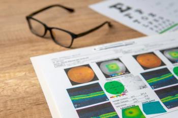
Diabetic retinopathy clinical pearl pictorial
Trace the path of diabetic retinopathy disease via images.
Retinal (macular) edema is caused by the breakdown of the blood-retinal barrier, resulting in leakage of serum, lipids and protein.
On clinical examination, edema may be difficult to see, but vascular changes such as microaneurysms and hard lipids known as exudates are often better visible (Figure 1A).
On optical coherence tomography (OCT), edema results in variable increases in the retinal or macular thickness, and fluid is often represented as hypo-reflective pockets (Figure 1B).
Following successful treatment with anti-vascular endothelial growth factor (VEGF) injections and/or laser photocoagulation, an increase in hard exudates may initially be noted as serum dries out while hard lipid residue forms (Figures 2A and 2B).
Once the leakage stops, it may take several months for the hard exudates—seen as intraretinal hyper-reflective deposits on the OCT—to slowly absorb (Figure 3).
Occasionally, the solidifying serum can be seen in mid-phase from liquid to hard deposits. This can be referred to as “soft exudate,” not to be confused with the use of the same term to describe cotton-wool spots. Clinically, “soft exudate” has a creamy color (Figure 4A); on OCT it has a semi hyper-reflective presentation (Figure 4B). After treatment, this material turns to hard exudate and dissipates over time (Figures 5 and 6).
Neglecting continued care of the disease can result in significant worsening of these findings and patients prognosis (Figure 7).
The patient depicted here failed to return for continued treatment for several months, resulting in increased severity of exudative retinopathy.
Articles in this issue
about 5 years ago
Quiz: Treatments for presbyopia coming soonabout 5 years ago
Quiz Answers: Treatments for presbyopia coming soonabout 5 years ago
When to lease and when to own office spaceabout 5 years ago
Vision rehabilitation of patients with strokeabout 5 years ago
Treatments for presbyopia coming soonabout 5 years ago
How diabetes affects COVID-19about 5 years ago
Novel uses of technology for systemic diseaseabout 5 years ago
Know the pros and cons of outsourcing billingabout 5 years ago
5 communication strategies for dry eye patientsNewsletter
Want more insights like this? Subscribe to Optometry Times and get clinical pearls and practice tips delivered straight to your inbox.


