
|Articles|April 14, 2021
- April digital edition 2021
- Volume 13
- Issue 4
Investigational agent aims to eradicate Demodex mites
Author(s)Milton M. Hom, OD, FAAO, FACAAI (Sc)
An FDA-approved drop would be a game changer in the treatment of Demodex blepharitis
Advertisement
OTC treatments for Demodex are available, but many are cumbersome and uncomfor table. No approved drugs are currently on the market. Effective treatments are needed because Demodex mites are the most common ectoparasite found in humans.1
Although these mites are common in small numbers, an overpopulation will infest the eyelash follicles, meibomian glands, and sebaceous glands.2 Their presence (Figure 1) has been implicated as a cause of chronic blepharitis, an ocular inflammation that involves primarily the eyelid margin.3-5 One meta-analysis of almost 5000 patients revealed that Demodex is implicated in 45% of blepharitis cases.6 In the United States alone, this translates to approximately 9 million individuals.
Despite the impact that Demodex blepharitis has on vast numbers of patients, no FDA-approved treatments exist. If left unmanaged, Demodex blepharitis may lead to tear film instability with fluctuating and blurred vision, lid and lash abnormalities, inflammation of the conjunctiva and surrounding skin, suboptimal surgical outcomes, contact lens intolerance and reduced wear time, noticeable eye and eyelid redness, and lower patient quality of life.3,7-10
Demodex clinical signs
Practitioners can easily see clinical signs of Demodex through a slit lamp. Demodex blepharitis can be diagnosed when collarettes, a pathognomonic sign of infestation,5,7,11 are observed (Figure 2). As mites scratch and feed on the skin, the partially digested epithelial cells, keratin, mite waste, and eggs combine to form collarettes.7,11 These collarettes are typically found at the base of the lash but can migrate up as the hair shaft grows.11
Demodex infestation leads to disease in 3 main ways. The first is mechanical, when overcrowded mites scrape the epithelial cell lining with their claws and lay eggs in the follicle, causing follicular distension, misdirected lashes, madarosis, and irritation. Dead mites and collarettes obstruct the hair follicle opening, leading to inflammation. The second way the mites cause disease is by excreting digestive enzymes as they feed and by exuding digestive waste when they die, leading to inflammation, hyperemia, irritation, and epithelial hyperplasia. Disease also occurs because the bacteria living on the mite’s surface or in its gut cause inflammation of the surrounding ocular tissues.
Clinical data overview
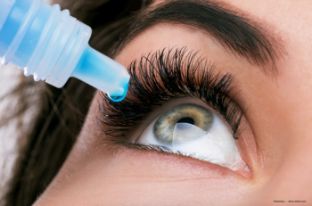
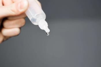
Currently, no FDA-approved therapy is indicated for Demodex blepharitis. Soon, however, eye care practitioners may have a new topical ophthalmic drop that can eliminate the mite by causing paralysis and death of the parasite. TP-03 (lotilaner 0.25%; Tarsus Pharmaceuticals) is an investigational agent that has shown promise in eradicating Demodex by inhibition of parasite-specific γ-aminobutyric acid chloride channels.
To date, 4 phase 2 clinical trials have revealed the agent to be well tolerated, safe, and effective. Specifically, results of the company’s Mars and Jupiter phase 2 studies have shown TP-03 is well tolerated and effective at reducing collarettes and Demodex density after treatment for 28 days, and the effect is maintained through at least 90 days.12,13 The 2 studies evaluated 75 patients, showing statistically significant decreases in collarettes and mite density as early as day 14 of treatment. No treatment-related adverse effects (AEs) were observed, and patients reported the drop to be comfortable.
These results were further validated in the phase 2a Io and phase 2b Europa studies that included a total of 72 patients.14 In the phase 2a Io, treatment with TP-03 was effective at achieving the primary and secondary end points, respectively, of collarette cure in 72% of participants and mite eradication in 78% of participants at day 42. In the phase 2b Europa trial, statistically significant results were achieved for the primary end point of collarette cure by 80% of participants on TP-03 compared with 16% on vehicle (P<.001) at day 42, and the secondary end point of mite eradication by 73% of participants on TP-03 compared with 21% on vehicle (P=.003) at day 42.
TP-03 was well tolerated, and no serious AEs or treatment discontinuations due to AEs were reported in either study. Participants in the Europa trial rated the administration of the eye drops as “neither comfortable nor uncomfortable,” “comfortable,” or “very comfortable” 87% of the time.
Jupiter study results
I recently presented the results of the phase 2 Jupiter study during the virtual Optometry’s Meeting in June 2020.13 A total of 60 patients were enrolled in the study, 30 in the active arm and 30 in the vehicle control arm. Participants had a mean age of 60.6 years, 41 (68.3%) were women, and all were Hispanic. The 2-arm, parallel study was conducted at a single site in Mexico City, Mexico, and participants were randomized 1:1 to the active or vehicle control study arms.
To be included, patients were required to have, in at least 1 eye, more than 10 collarettes present on the upper lid; mild to severe lid margin erythema; and an average mite density of 1.5 mites or more per lash (upper and lower eyelids combined).
Investigators graded collarettes at the slit lamp. In potential participants who met all other entry criteria, investigators determined mite density by epilating 4 to 6 lashes from each eye. Mites were counted by viewing the lashes under a microscope. Mite density was computed as the total number of mites counted for each eye divided by the total number of lashes. Participants administered 1 drop of their assigned treatment in each eye, twice daily, for 28 days and were assessed at days 7, 14, 28, 60, and 90. Efficacy was determined by the decrease in collarettes and the decrease in mite density. Safety was determined by assessing treatment-related AEs as well as evaluating any changes in visual acuity, intraocular pressure (IOP) and slit lamp biomicroscopy findings, and corneal staining.
This study demonstrated that use of TP-03 was well tolerated during the 28-day treatment and effective in reducing collarettes and Demodex density through 90 days in participants with Demodex blepharitis. The change in collarette grade demonstrated statistically significant decreases for both eyes, upper and lower eyelid margins, compared with the control arm, with decreases persisting for at least 2 months following treatment. Investigators observed statistically significant decreases in mean mite density compared with vehicle control in both eyes beginning at day 28, with decreases persisting for an additional 2 months following treatment.
No treatment-related AEs were reported, and there were no clinically significant changes in visual acuity or IOP. Clinically significant changes in slit lamp biomicroscopy findings, primarily related to corneal staining in 2 participants in the active group and 6 participants in the control group, were observed. Two participants in the control group experienced mild conjunctival hyperemia in both eyes; this resolved without treatment. Further, 90% of patients reported the drop to be neutral to comfortable.
My experience
I believe that in anterior segment disease, Demodex is the most underdiagnosed, underappreciated, and undertreated ocular surface disease. Demodex is quite common in my practice, with approximately 25% of patients presenting with an infestation. Most current therapies clean up the collarettes or cylindrical dandruff that results from an infestation. I use tea tree oil wipes on these patients, with both in-office treatment and take-home wipes for more severe cases. Although the oil can kill the mites, it is extremely uncomfortable and harsh on the ocular surface. In fact, if a clinician misses the lid, tea tree oil can cause a corneal burn. Results of a study showed that terpinen-4-ol or T4O—the tea tree oil component that has shown the most efficacy in killing Demodex mites—is toxic to human meibomian gland epithelial cells in vitro.15 This was shown even at levels 10-fold to 100-fold lower than the demodicidal concentration used to kill the mites.
Fishman and colleagues have reported on the use of intense pulsed light (IPL) to kill Demodex in vitro and in real time.16 They suggest that IPL application at settings identical to those used to treat meibomian gland disease can destroy the mite.
Although not yet studied in combination with other treatments, TP-03 could be used as part of a multipronged approach to ocular surface disease. I believe that it may be possible that a Demodex infestation can be an underlying cause of dry eye disease and meibomian gland disease. Of course, an infestation can also be present in the absence of other ocular surface disease.
Conclusion
TP-03, if approved, would be the first therapeutic available specifically for the treatment of Demodex blepharitis. Data to date are encouraging.
Tarsus announced it has begun patient enrollment and treatment in its Saturn-1 phase 2b/3 pivotal trial. Saturn-1 is a larger, multicenter trial with the same end points as those of Europa. Enrollment in a second pivotal trial, Saturn-2, is expected to begin in 2021.
Many times, I have been asked whether there will ever be a drug that will treat this parasite. I am now hopeful there is one on the horizon.
References
1. English FP. Demodex folliculorum and oedema of the eyelash. Br J Ophthalmol. 1971;55(11):742-746. doi:10.1136/ bjo.55.11.742
2. Rufli T, Mumcuoglu Y. The hair follicle mites Demodex folliculorum and Demodex brevis: biology and medical importance. a review. Dermatologica. 1981;162(1):1-11. doi: 10.1159/000250228
3. Amescua G, Akpek EK, Farid M, et al; American Academy of Ophthalmology Preferred Practice Pattern Cornea and External Disease Panel. Blepharitis preferred practice pattern. Ophthalmology. 2019;126(1):PP56-PP93. doi:10.1016/j. ophtha.2018.10.019
4. Coston TO. Demodex folliculorum blepharitis. Trans Am Ophthalmol Soc. 1967;65:361-392.
5. Gao YY, Di Pascuale MA, Li W, et al. High prevalence of Demodex in eyelashes with cylindrical dandruff. Invest Opthalmol Vis Sci. 2005;46(9):3089-3094. doi:10.1167/ iovs.05-0275
6. Zhao YE, Wu LP, Hu L, Xu JR. Association of blepharitis with Demodex: a meta-analysis. Ophthalmic Epidemiol. 2012;19(2):95-102. doi:10.3109/09286586.2011.642052
7. Fromstein SR, Harthan JS, Patel J, Opitz DL. Demodex blepharitis: clinical perspectives. Clin Optom (Auckl). 2018;10:57-63. doi:10.2147/OPTO.S142708
8. Lemp MA, Nichols KK. Blepharitis in the United States 2009: a survey-based perspective on prevalence and disease. Ocul Surf. 2009;7(suppl 2):S1-S14. doi:10.1016/s1542- 0124(12)70620-1
9. Eye and vision conditions. American Optometric Association. Accessed March 10, 2021. https://www.aoa.org/healthy-eyes/ eye-and-vision-conditions?sso=y
10. Treat blepharitis preoperatively for optimal cataract surgery results. Primary Care Optometry News. November 1, 2010. Accessed March 10, 2021. https://www.healio.com/news/ optometry/20120225/treat-blepharitis-preoperatively-for-optimal-cataract-surgery-results#:~:text=According%20 to%20Dr.,Swartz%20said
11. Nicholls SG, Oakley CL, Tan A, Vote BJ. Demodex species in human ocular disease: new clinicopathological aspects. Int Ophthalmol. 2017;37(1):303-312. doi:10.1007/s10792-016- 0249-9
12. Quiroz-Mercado H, Ramos-Betancourt N, Corredor-Ortega C, et al. Pilot study to evaluate the safety and efficacy of TP-03 for the treatment of blepharitis due to Demodex infestation (Mars study). Invest Ophthalmol Vis Sci. 2020;61(7): 2984.
13. Hom MH, Ceballos JC, Massaro-Corredor M, et al. Randomized controlled trial to evaluate the safety and efficacy of TP-03 for the treatment of blepharitis due to Demodex infestation (Jupiter study). Poster presented at: 123rd Annual American Optometric Association’s Optometry’s Meeting; June 24-28, 2020; virtual.
14. Tarsus releases data from Io and Europa trials for TP-03 to treat Demodex blepharitis and begins enrollment and treatment in phase 2b/3 Saturn-1 trial. News release. October 6, 2020. Accessed March 10, 2021. https://www.prnewswire.com/ news-releases/tarsus-releases-data-from-io-and-europa-trials-for-tp-03-to-treat-demodex-blepharitis-and-begins-enrollment-and-treatment-in-phase-2b3-saturn-1-trial-301146578.html
15. Chen D, Wang J, Sullivan DA, Kam WR, Liu Y. Effects of terpinen-4-ol on meibomian gland epithelial cells in vitro. Cornea. 2020;39(12):1541-1546. doi:10.1097/ ICO.0000000000002506
16. Fishman HA, Periman LM, Shah AA. Real-time video microscopy of in vitro Demodex death by intense pulsed light. Photobiomodul Photomed Laser Surg. 2020;38(8):472-476. doi: 10.1089/photob.2019.4737
Articles in this issue
about 5 years ago
The coming presbyopia revolutionabout 5 years ago
Biologic medications: The basics that every OD should knowabout 5 years ago
How to fit scleral lenses with confidence and cautionabout 5 years ago
Quiz: How to fit scleral lenses with confidence and cautionabout 5 years ago
Where golf and optometry meetings intersectabout 5 years ago
News updates April 2021about 5 years ago
5 glaucoma management mythsabout 5 years ago
Treating sight-threatening retinopathy using OCTAAdvertisement
Related Content
Advertisement


Latest CME


Advertisement
Advertisement
Trending on Optometry Times - Clinical News & Expert Optometrist Insights
1
AOA 2026: AREDS2 and AREDS2+B vitamin complex implications for AMD
2
Thea Pharma launches Zolymbus in the US as preservative-free bimatoprost gel
3
AOA 2026: The latest updates in keratoconus treatment advancements
4
First EDOF contact lens improves intermediate, near visual acuity in presbyopes, study finds
5