Study finds electroretinography/pupillometry as strongest predictor of DR progression, vision-threatening complications
Data used in the post hoc analysis were from 56 parameters from 4 testing modalities obtained in a multicenter US trial (NCT03197870).
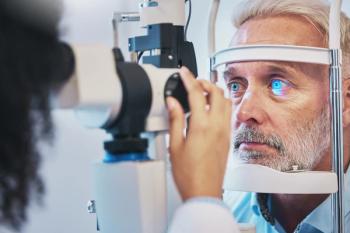
A recent longitudinal prospective study revealed that electroretinography (ERG)/pupillometry outperforms structural imaging in identifying patients at the highest risk of progressing to vision-threatening complications over 1 year. The study was published in Ophthalmology Science and provided a post hoc analysis of 56 parameters from 4 testing modalities obtained in a multicenter US trial (NCT03197870), according to a news release.1 Eighteen of these parameters were from the RETeval device’s diabetic retinopathy (DR) Assessment protocol, graded by manufacturer LKC Technologies, which was found to be the strongest predictor of DR progression.2
“The DR score, based on a combination of ERG and pupillometry, offered the best predictive capability.… These results suggest it is possible to improve the DR staging system, which in turn may enable better allocation of health care resources,” the study authors, led by C. Quentin Davis, PhD, of LKC Technologies, stated.
“This publication is a significant advancement in the quest to identify high-risk patients earlier and improve clinical outcomes,” said Dina Dubey, CEO of LKC Technologies, in the release. “The prospective study included patients with moderate to severe non-proliferative diabetic retinopathy (NPDR) and no center-involved DME at baseline and it revealed that a single number – known as the DR Score – is the most powerful predictor of progression from NPDR to vision-threatening complications, including proliferative diabetic retinopathy, diabetic macular edema, or treatment.”
The trial used in the post hoc analysis for data collection was the prospective, double-masked, randomized, placebo-controlled TIME-2b study that evaluated the safety and efficacy of 48 weeks of subcutaneous AKB-9778 (a Tie2 activator) in patients with moderate to severe NPDR. Every 12 weeks, patients underwent a comprehensive ocular assessment including a dilated slit lamp biomicroscopy, intraocular pressure, dilated indirect ophthalmoscopy, spectral-domain OCT, and 7-field FP. FA was conducted at weeks 0, 24, and 48. ERG/pupillometry, OCTA, and UWF-FA were performed using commercially available equipment at a subset of the 44 sites included in the study based on availability.2
The DR Score of 26.9 demonstrated a relative risk of 5.6 for progression to vision-threatening conditions (P<.0001), surpassing other predictors including ultra-widefield fluorescein angiography (UWF-FA), optical coherence tomography angiography (OCTA), and fundus photography (FP). Comparatively, the UWF-FA total ischemia index ≥ 0.125 had a relative risk of 5.3 (P<.0001), the OCTA foveal avascular zone area ≥ 0.295 mm2 had a relative risk of 3.6 (P<.05), and the fundus photograph-based diabetic retinopathy severity scale ≥ 27 had a relative risk of 2.1 (P<.05), each still proving statistical significance.1
“There is mounting evidence that DR's effects on retinal neurons occur earlier and can progress somewhat independently of changes in retinal vasculature,” the study authors stated. “Thus, it is likely that combining measures of neuronal function with structural vascular information should enhance prediction of DR progression.
“This research suggests that the ERG provides value in defining how DR is staged and monitored, supporting mounting evidence that functional changes in the retina can precede observable vascular abnormalities,” said Mitchell Brigell, PhD, one of the study authors, in the release. “The findings clearly show the value of integrating objective functional testing into the management of diabetic retinopathy. This is also reflected in the inclusion of ERG in the American Academy of Ophthalmology’s Preferred Practice Pattern Guidelines for Diabetic Retinopathy.”
Limitations of the study include its unequal number of subjects across modalities, with fewer eyes tested with OCTA, UWF-FA, and ERG/pupillometry than FP due to equipment availability, in addition to the binary classification of high or low risk, according to study authors.2 “Thus, performance differences may reflect sampling variation, although the reported uncertainties account for differences in sample size,” the authors stated.
References:
Study identifies RETeval DR Score as the strongest predictor of diabetic retinopathy progression. LKC Technologies. August 4, 2025. Accessed August 12, 2025.
Davis CQ, Waheed NK, Brigell M. Predicting progression to vision-threatening complications in diabetic retinopathy. Ophthalmol Sci. 2025;5(6). doi:10.1016/j.xops.2025.100859
Newsletter
Want more insights like this? Subscribe to Optometry Times and get clinical pearls and practice tips delivered straight to your inbox.


