
|Articles|January 23, 2019
Blog: Modern-day techniques for diagnosing dry eye disease
Author(s)Tracy Schroeder Swartz, OD, MS, FAAO
Advertisement
The views expressed here belong to the author. They do not necessarily represent the views of Optometry Times or UBM.
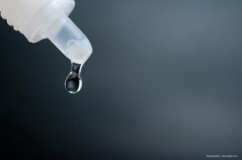
Remember when ODs diagnosed dry eye using a slit lamp and treated with artificial tears? I think it was back when we refracted with loose lenses and panicked if a soft contact lens dropped on the floor.
We used paper charts, wrote on real paper, and mailed letters in envelopes with stamps we had to lick to get them to stick. Life was so simple then.
ODs today have computerized phoropters, daily disposable contact lenses, and electronic medical records (EMR). We rarely talk to a pharmacist because we send prescriptions over the Internet. Dry eye diagnosis and treatment have become increasingly complex in this not-so-simple age.
Our understanding of ocular surface disease has expanded to include highly sophisticated testing. Patients are no longer told to “use artificial tears and see me in a year.” Evaporative dry eye has refocused dry eye treatment to the lids.
Here is a list of the modern testing techniques ODs use for
Vital dye staining of cornea/lids/conjunctivae
Sodium fluorescein dye for epithelial integrity is the mainstay for corneal staining. It permeates the intercellular space associated with epithelial cellular disruption and is also beneficial for break-up time assessment and epithelial basement membrane dystrophy (EBMD) evaluation.
Lissamine green is preferred for staining the conjunctivae. Lissamine green stains dead and dying cells on the cornea and bulbar conjunctiva. Its affinity for conjunctivae facilitates diagnosis of lid wiper epitheliopathy (LWE). It is also tolerated better than rose bengal.
Lid expression
Lid expression can be performed diagnostically using a finger, swab, or tool. Gentle pressure applied to the lid margin to assess the meibomian gland function is newly recommended to assess lid health.
You may be surprised how many glands look relatively normal until you apply pressure to find out nothing is expressed. Paddles are used to therapeutically treat meibomian gland dysfunction (MGD) by evacuating the glands more vigorously.
Meibography
Using meibography with infrared imaging allows assessment of gland architecture and identification of gland loss. Other techniques less commonly used for meibography include ocular coherence tomography (OCT) and laser confocal microscopy.
Infrared systems may be added to the slit lamp or be stand-alone systems. An OD may be able to bill for anterior segment photography (CPT code 92285) for this procedure.
Blink rate assessment, quality of blink
Patients with ocular surface disease have trouble holding their eyes open without blinking, and may blink incompletely.1
Lipid layer assessment
Using interferometry in lipid layer assessment investigates the stability of the lipid layer and likelihood of evaporative dry eye. Measurement involves assessment of the interference pattern produced by the inner and outer surfaces of the lipid layer in tear film.
This has been shown to differentiate between patients with normal eyes and those with dry eyes because patients with dry eyes tend to have thinner lipid layers.2 However, these systems are more expensive.
Topography and aberrometry
Topography and aberrometry become more irregular with ocular surface disease. This increases higher-order aberrations on both whole eye wavefront and corneal wavefront due to poor tear film and corneal abnormalities.
Tear break-up time
Performing tear break-up time using sodium fluorescein dye (FTBUT) has long been done but is now performed using computer imaging to time the surface images and blinking.
This test is now referred to as non-invasive tear break-up time (NIBUT). Various systems allow this measurement, and further investigations will provide a better understanding of the usefulness of NIBUT patterns in addition to time measurements.
Tear meniscus measurement
This is used to assess secretion rate and film stability. It is performed with computers using infrared or visible light as well as OCT.
Matrix metalloproteinase 9 (MMP-9) measurement
This measurement is used in office testing to indicate the presence of proteins that stimulate the immune response and is associated with ocular surface disease progression.
Significant increases in MMP-9 results were found in women patients with autoimmune diseases-especially Sjögren syndrome-and thyroid disease.
References:
1. White R, Rodriguez J, Lane KJ, Johnston P, Angjeli E, Abelson MB. Blink patterns in normal and dry eye subjects; beyond blink rate. Invest Opthalmol Vis Sci. 2010 Apr;(51):3366.
2. Hwang HS, Kim EC, Kim MS. Novel tear interferometer made of paper for lipid layer evaluation. Cornea. 2014 Aug;33(8):826-31.
3. Messmer EM, von Lindenfels V, Garbe A, Kampik A. Matrix metalloproteinase 9 testing in dry eye disease using a commercially available point-of-care immunoassay. Ophthalmology. 2016 Nov;123(11):2300-2308.
Advertisement
Related Content
Advertisement


Latest CME


Advertisement
Advertisement
Trending on Optometry Times - Clinical News & Expert Optometrist Insights
1
3D-printed contact lenses offer patient-specific fit in 20 minutes
2
Atropine, aspherical lenses enter trial for childhood myopia
3
Classifying Dry Eye by Root Cause: A Step Toward Precision Medicine
4
First patient enrolled in pilot cataract trial assessing lanosterol eye drop ZOC2017217
5