
|Articles|November 1, 2009
Changes in the profession are moving at fast pace
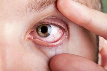
Glaucoma technology is swiftly evolving.
Advertisement
As technology improved, so did the ability to assess visual fields in a faster, more reliable way. Fundus photography enhanced the clinician's ability to detect change by being able to compare either slides or Polaroid pictures of an optic nerve over time. The digital revolution gave us the ability to capture photos of the optic nerve and analyze them in ways we could never have done in the past. Additionally, with the press of a button, you could share these images with a colleague across town or across the continent.
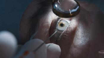
Optical coherence tomography (OCT) provides the practitioner with a virtual biopsy of the retinal tissue, including the optic nerve and retinal nerve fiber layer thickness. Additionally, the newest generation OCTs offer the ability to acquire significantly more information than their time domain predecessors did and in a fraction of the time. This newest generation, often referred to as spectral domain or Fourier domain, allows an analysis of the ganglion cell complex, which represents the nerve fiber layer, ganglion cell layer, and inner plexiform layer in the macular area, which may detect glaucomatous changes earlier.
Prior to the publication of the Ocular Hypertension Treatment Study (OHTS), central corneal thickness was not routinely measured in cases of suspected glaucoma and patients with glaucoma. Since this breakthrough publication, OHTS has become the standard of care for treating patients with glaucoma. Additionally the data reported in the study has given rise to risk calculators that can be used clinically to assess an ocular hypertensive's risk of developing glaucoma over the next 5 years.
With all the technological advances that are available to help manage this complex, devastating disease, deciding which is best can be overwhelming. In reality, there is no wrong answer. Each instrument has its strengths and a place in the practice. Through careful research and analysis, you can find which device is right for your patient base.
Imaging technology is becoming a standard in practices across the United States. Thousands of practitioners are embracing this technology, thereby enhancing the care that they deliver to their patients.
Do the research, do the analysis, and-most importantly-don't be left behind.
David L. Kading OD, FAAO, practices in Seattle, WA. Dr. Kading's practice specializes in anterior segment disease and contact lens fitting. He can be reached at
.
Mile Brujic, OD, is a partner of a four location optometric practice in northwest Ohio. Dr. Brujic may be contacted at
.
Newsletter
Want more insights like this? Subscribe to Optometry Times and get clinical pearls and practice tips delivered straight to your inbox.
Advertisement
Related Content
Advertisement


Advertisement
Advertisement
Trending on Optometry Times - Clinical News & Expert Optometrist Insights
1
Dry eye and macular degeneration: A convergence of aging, inflammation, and telomere biology
2
Dry eye an area of prime interest to ODs and MDs in 2025
3
Detecting glaucoma progression based on multiple retinal layers' thickness
4
Refractive surgery for myopia vs presbyopia: Tailoring the approach for the individual
5