Demodex “tails”
A review of Demodex mites.

")
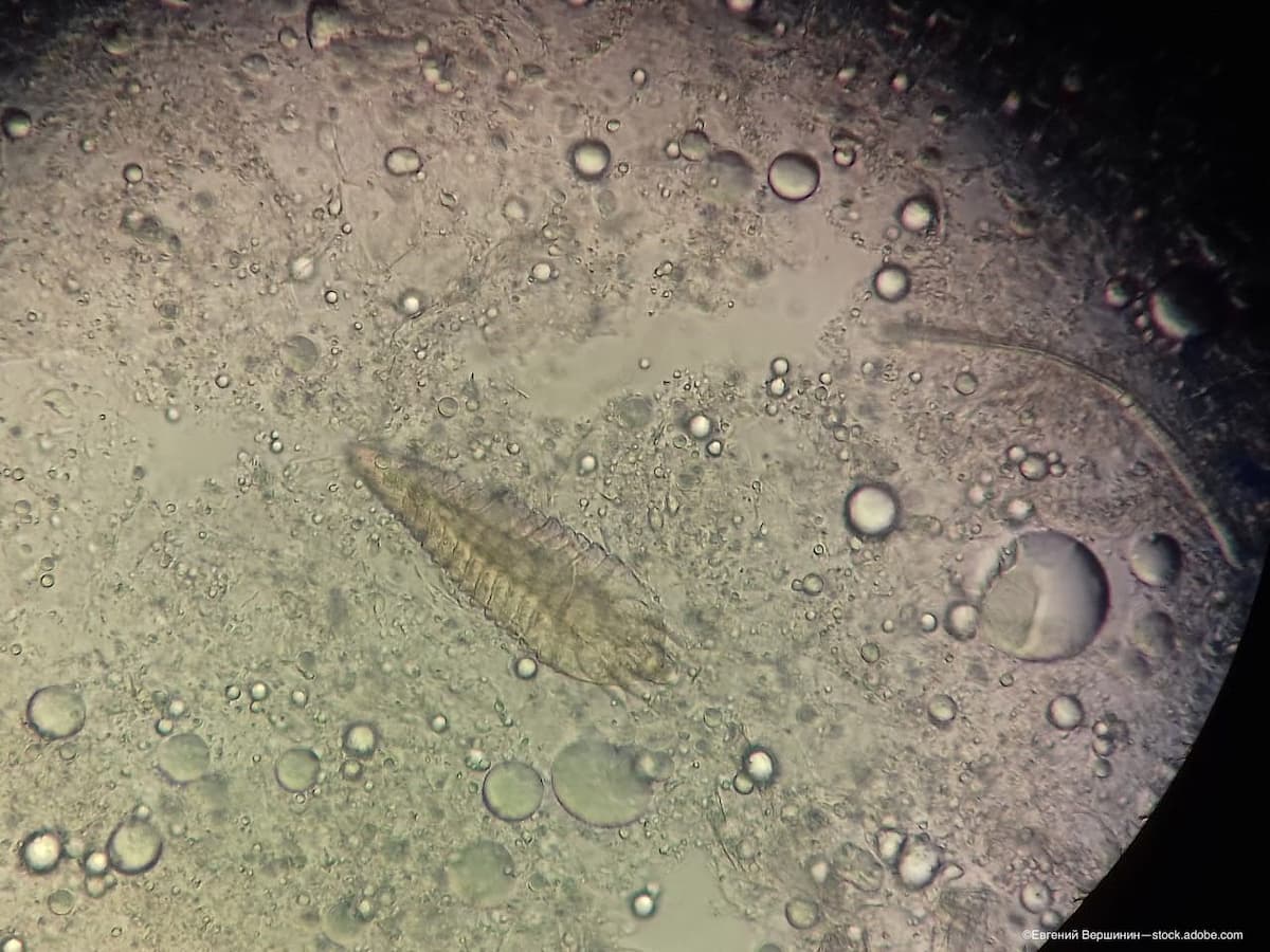
Demodex folliculorum is the most common ectoparasite of man; it does not leave its human host. (Adobe Stock / Евгений Вершинин)
More than 100 different species of follicular mites have been morphologically described and studied in mammals ranging from marsupials to placental creatures such as armadillos, bats, pigs, dogs, rodents, and primates.1 Interest in the Demodex mite (class: Archnida; family: Demodidicidae; genus: Demodex; species: Demodex folliculorum) as a contributor to blepharitis has waxed and waned since it was first reported in the eyelash follicle in the mid-1800s. With expanded investigation and novel study techniques, information on these mites is advancing, giving us a better understanding of the organism.
Demodex folliculorum is the most common ectoparasite of man; it does not leave its human host. It aggregates in groups in the infundibular portion of hair follicles, most numerous in the wings of the nose, the ear canal, and the nipples. It has been isolated in other hair-bearing parts of the body, including but not limited to the face, chest, pubis, armpits, scalp, ear canal, eyebrow, and eyelash follicles. It is proposed that Demodex transmission is horizontal from mother to offspring—the increased temperature and moisture levels at the nipple during breastfeeding facilitate transfer of the mites from mother to offspring.1
Demodex life cycle
The life cycle of Demodex from egg to nymph to adult is roughly 2 weeks, with adult mites living for an additional 1 to 2 weeks. The genome of the Demodex mite encodes for 9707 proteins.1 The mite is 0.2 to 0.3 mm long with a semitransparent, vermiform, elongated body consisting of 2 fused, translucent, chitinous segments (head/tail) with a head-to-body ratio of 1:2. The front of the body is referred to as the gnathosoma; the posterior, cigar-shaped portion is the opisthosoma. The mouthparts (gnathosoma) of Demodex include a clawed palpus (feeler attached to mouthparts), a round oral opening (1211 nm), and a sharp oral needle (300 nm). The adult mite has 4 pairs of bud-type walking legs (podosoma) approximately 15 µm long on either side of its body with 3 mobile segments and 1 fixed segment (podomeres), and can locomote at 8 to 16 mm/h. There are 2 to 3 tined claws and spurs on each leg to assist mobility.
The Demodex mite has the simplest brain observed in Acari, but it occupies a large volume in relation to total body size.2 Part of the brain is around and below the legs.
The Demodex mite has a pair of dorsoanterior photosensitive organs known as “supraconal spines,” and the “eyes” contain a handful of ciliary photoreceptors protruding from a large cell containing pigment. An opsin-like protein present in Demodex initiates the transduction cascade necessary in the detection of light and possess an almost complete circadian rhythm pathway. In accord with nocturnal habits, Demodex lacks genes for UV protection.1
As far as digestion, Demodex have an incomplete gut, with the hindgut being a fingerlike minute tube that opens to the outside at approximately one-third of the posterior terminus of the opisthosoma. It is unlikely that the waste of Demodex is contained until death.1
The sexes are separate in mites; males have a pair of testes in the midregion of the body, each connected to the gonopore by a vas deferens, and a chitinous penis; females have a single ovary connected to the gonopore by an oviduct, as well as a seminal receptacle for the storage of sperm. The male genital orifice is placed dorsal, between a second pair of legs; the vulva extends ventrally at the level of the fourth pair of legs. Females of Demodex folliculorum are larger and rounder than males. Fertilization is internal.3 The arrow-shaped eggs are laid in the substrate, or wherever the mite happens to live. The eggs hatch, with the next stage being the 6-legged larvae. The female lays 20 to 24 eggs, and their size reaches 50 to 60 μm, from which the larvae hatch molting and morphing into nymphs (6 legs) and further into adults (8 legs).4
Conclusion


In the eyelash follicle, Demodex can be identified by direct examination at the slit lamp at 40× magnification and appear as translucent rodlike sticks extending above the skin surface close to the eyelash follicles.5 Likewise, the lash can be rotated6 or tractioned7 within the follicles to coax Demodex out of the follicle. Demodex can be viewed by in vivo confocal microscopy8 or may be visualized by light microscope examination of an epilated eyelash.9
Demodex mites are significant, as they are ubiquitous in humans and play a role in dermatoses. Continued discovery will help us understand the conundrum of Demodex as host-injuring obligate parasite to an obligate symbiont.1
References
1. Smith G, Manzano Marín A, Reyes-Prieto M, et al. Human follicular mites: ectoparasites becoming symbionts. Mol Biol Evol. Published online June 21, 2022. doi:10.1093/molbev/msac125
2. Beutel RG, Pohl H, Hünefeld F. Strepsipteran brains and effects of miniaturization (Insecta). Arthropod Struct Dev. 2005;34(3):301-313. doi:10.1016/j.asd.2005.03.001
3. Rush A. Demodex folliculorum. Animal Diversity Web. Accessed May 23, 2023. https://animaldiversity.org/accounts/Demodex_folliculorum—Demodex/
4. Lacey N, Kavanagh K, Tseng SC. Under the lash: Demodex mites in human diseases. Biochem (Lond). 2009;31(4):2-6.
5. Gao YY, Wang T, Jiang YT, et al. Should ocular Demodex be checked and treated in refractory keratitis patients without blepharitis? Int J Ophthalmol. 2023;16(2):201-207. doi:10.18240/ijo.2023.02.05
6. Mastrota KM. Method to identify Demodex in the eyelash follicle without epilation. Optom Vis Sci. 2013;90(6):e172-e174. doi:10.1097/OPX.0b013e318294c2c0
7. Muntz A, Purslow C, Wolffsohn JS, Craig JP. Improved Demodex diagnosis in the clinical setting using a novel in situ technique. Cont Lens Anterior Eye. 2020;43(4):345-349. doi:10.1016/j.clae.2019.11.009
8. Liang H, Randon M, Michee S, Tahiri R, Labbe A, Baudouin C. In vivo confocal microscopy evaluation of ocular and cutaneous alterations in patients with rosacea. Br J Ophthalmol. 2017;101(3):268-274. doi:10.1136/bjophthalmol-2015-308110
9. Gao YY, Di Pascuale MA, Li W, et al. High prevalence of Demodex in eyelashes with cylindrical dandruff. Invest Ophthalmol Vis Sci. 2005;46(9):3089-3094. doi:10.1167/iovs.05-0275

 on blue background, medically 3D illustration (Adobe Stock / Axel Kock)")
")


")
")
")
")