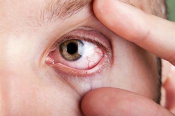
Study explores eye health after COVID-19 recovery through the choroidal vascularity index
A total of 43 patients who recovered from the SARS-CoV-2 infection with mild pneumonia were included along with 45 healthy individuals.
Researchers from Istanbul, Turkey, conducted a study that found that the choroidal vascularity index (CVI) can shed light on the choroidal vascular physiology in patients who had had mild
A total of 43 patients who recovered from the SARS-CoV-2 infection with mild pneumonia (group 1, COVID group) were included along with 45 healthy individuals (group 2, healthy control group).
All fully recovered patients were evaluated 6 months after the pneumonia resolved. The investigators measured the choroidal structures using enhanced depth imaging optical coherence tomography (EDI-OCT). The primary measurement of interest was the CVI, which the investigators defined as the ratio of the luminal area to the total choroidal area.
The patients who comprised group 1 (COVID group) had significantly higher values compared to the healthy group 2 controls in the mean total choroidal area, the stromal area, and the luminal area. There was no difference in the CVI between the 2 groups (p = 0.080).
Based on their findings, the investigators concluded that the CVI can reveal the choroidal vascular physiology in patients who have fully recovered from COVID-19 pneumonia. The EDI-OCT technology can be used to evaluate choroidal vascular alterations and thereby serve as a non-invasive indicator for early vascular impairment following SARS-CoV-2 infection.
Reference:
Toprak M, Kesim E, Karasu B, Celebi ARC. Choroidal vascularity index findings in patients recovered from mild course COVID-19 pneumonia.Int Ophthalmol. 2025;45:84. https://doi.org/10.1007/s10792-025-03450-4
Newsletter
Want more insights like this? Subscribe to Optometry Times and get clinical pearls and practice tips delivered straight to your inbox.


