
|Articles|March 2, 2021
- March digital edition 2021
- Volume 13
- Issue 3
Treat what you are able when diagnosis is tricky
Author(s)Benjamin P. Casella, OD, FAAO
Advertisement
Every now and then I am reminded that sometimes things actually do make sense. Not long ago, a 20-year-old Caucasian man presented with pain behind his eyes.
He stated that the pain had been off and on for several months and that he had experienced limited relief with over-the-counter (OTC), nonsteroidal, anti-inflammatory pills. He did not associate the pain with any particular activity such as reading or computer work, and said his vision seemed fine at distance and near with no correction. He also denied worsening of the pain with changing his head posture and denied waking up with it at night.
He described the pain as a dull ache behind and around his eyes, which could last from a few seconds to several minutes. He said that laying down in the dark seemed to help a bit. He had seen his primary-care physician about this and underwent neuroimaging. He did not know what type of test was performed, but he could confirm that it was a brain scan and was clear.
Testing
The patient’s medical history was remarkable for seasonal allergies for which he took OTC antihistamines, as needed. He had no family history of glaucoma. He had no other apparent health problems, took no other medications, and had no known medication allergies. He also stated that he ate a healthy diet, regularly exercised, and drank plenty of water.
Entering distance visual acuities unaided were 20/20 OD and OS. Pupil function was normal for each eye, and extraocular muscle testing showed a full range of motion for each eye. In addition, confrontational visual field testing showed all 4 quadrants intact for each eye. Color vision was also intact for each eye.
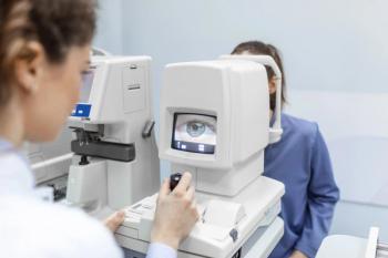
Manifest refraction showed a very low degree of simple hyperopia in each eye. Intraocular pressures (IOPs), measured by means of rebound tonometry, were 26 mm Hg in the right eye and 27 mm Hg in the left. IOPs measured by Goldmann applanation tonometry were 25 mm Hg in the right eye and 27 mm Hg in the left.
Anterior segment examination showed clear and quiet lids and lashes. Conjunctivas were clear and quiet, as were corneas. Both anterior chambers were deep and quiet with angles open to grade 3 via the von Herrick assessment method. Irises were intact and brown and frankly flat. Dilated fundus examination showed both retinas to be intact and flat. Blood vessels were dry and were of normal course and caliber.
Optic nerves were healthy, distinct, and well perfused with a cup-to-disc margin of 0.3 horizontally and vertically for each eye. Retinal nerve fiber assessment with the use of a pre-corneal lens and a red-free filter showed both retinal nerve fiber layers (RNFL) around the optic nerves to be robust without defects.
Follow-up
At this point, I explained to the patient that his IOPs were on the high side but that there was no frank evidence that they were causing him to have glaucoma. I invited him back in a couple of weeks for glaucoma-specific testing and asked him to bring the results of his neuroimaging with him (he had a follow-up visit with his primary-care doctor in the meantime).
Since the patient’s first visit was in the midmorning, I invited him back in the afternoon for his follow-up visit. At the follow-up visit, entering unaided distance visual acuities were unchanged at 20/20 for each eye. Pupils were still functioning well, as were extraocular muscles for each eye. Visual fields were intact for each eye, as well.
Anterior segment examination was normal and unchanged for each eye, and optic nerves were still distinct and perfused as viewed with a precorneal lens through undilated pupils. IOP measured by means of Goldmann applanation tonometry at 3:40 pm were 27 mm Hg in the right eye and 25 mm Hg in the left. A 24˚ threshold visual field study was unremarkable for each eye, and optical coherence tomography (OCT) studies showed a clear RNFL and a healthy and robust ganglion cell complex for each eye.
Central corneal thickness values were 578 µm for the right eye and 580 µm for the left. Gonioscopy revealed angles open to the ciliary body with a flat iris approach and mild pigmentation in all 4 quadrants for each eye. He brought with him the results of his neuroimaging study, and thankfully had had undergone a whole brain magnetic resonance imaging (MRI) with and without contrast, which was essentially clear. He reported having a couple of episodes of pain around his eyes since his first visit with me.
Treatment
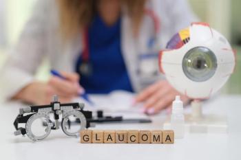
At this point, I informed the patient that he had high IOPs, which was a risk factor for glaucoma but no signs of the disease. However, with nothing else to treat, I thought perhaps his ocular hypertension could be causing him some pain. Maybe it was spiking up from time to time over the amounts I had measured. His pulse was 84 beats per minute and was regular. So, I prescribed timolol maleate 0.5% ophthalmic solution to be used as 1 drop in each eye every morning.
After educating him on the signs and symptoms of potential adverse effects of beta blocker, I invited him back in 1 month for another IOP check. At that visit, vision was still 20/20 in each eye, and pupils were normal. An undilated view of optic nerves showed distinct margins and good perfusion. IOPs were 18 mm Hg in each eye (in the midmorning).
The patient reported no episodes of pain with the timolol and no adverse effects of the medication since the initiation of therapy. I saw him for follow-up care 3 months later. His IOP was unchanged and the pain around his eyes had not yet returned.
In my experience, IOP needs to be pretty high (out of the 20s) to cause pain. That is one reason why so many people suffer from glaucoma before it is detected. In this case, I am thankful that just treating what I saw seems to have done the trick.
Articles in this issue
about 5 years ago
Vision rehabilitation of patients with traumatic brain injuryabout 5 years ago
Know risks and benefits of ocular steroid useabout 5 years ago
How to address dry eye in the challenging corneaabout 5 years ago
New data in anterior segment laser surgeryabout 5 years ago
Know 4 types of allergic eye diseaseabout 5 years ago
New therapy addresses dry eye flaresabout 5 years ago
COVID-19 & migraine: Patient impact & management tipsabout 5 years ago
Quiz: New data in anterior segment laser surgeryabout 5 years ago
How to prevent infection after LASIK or PRKAdvertisement
Related Content
Advertisement

Latest CME


Advertisement
Advertisement
Trending on Optometry Times - Clinical News & Expert Optometrist Insights
1
TearSolutions advances Lacripep into phase 2 following dual FDA designations in neurotrophic keratitis
2
Georgia university receives state funding for state's first college of optometry
3
Recent study findings may prove previous estimations of retinal cell communication, low-light vision
4
Exercise for the prevention and mitigation of eye disease
5