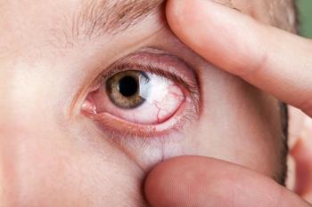
Zero in on differential diagnosis of uveitis
Developing a differential diagnosis for a patient with uveitis begins by identifying known risk factors for uveitic entities. Spend the time to take a thorough history. You’ll find that the huge number of possibilities has been trimmed to a manageable number, which can then be evaluated by direct examination and laboratory testing for an underlying disease in your uveitic patient.
Deloping a differential diagnosis for a patient with uveitis is often challenging, not only for the optometric physician, but for uveitis specialists as well. Patients affected with uveitis from both idiopathic and secondary etiologies present with a multitude of ocular and systemic signs and symptoms. It is in this grab bag of signs and symptoms that the diagnostician finds some clinical gems that assist him or her in finding the underlying etiology of the ocular inflammation.
Uveitis is estimated to account for 10% of visual impairment in the world today.1 It is also responsible for 30,000 new cases of blindness each year.1 Uveitis can occur in any age group, but affects those aged 20 to 44 years more commonly.
Figure 1. The ocular examination facilitates identification of keratic precipitates on the corneal endothelium and inside the anterior chamber.The incidence of uveitis in elderly patients may be higher than previously suspected, according to a recent study of adults 65 years of age and older.2 In this study, anterior uveitis was the most common form of uveitis encountered with an average of 243.6/100,000 persons per year. It is important to mention that in this age group, masquerade syndrome should always be considered as part of an ocular examination rule-out. Malignant tumors of the anterior segment ciliary body melanoma, may seed the anterior chamber with what appears to be inflammatory cells but, on close inspection, do not fit the diagnosis of uveitis.
Anterior uveitis has been reported to be the most common of the uveitic presentations to eyecare professionals (ECPs). It accounts for 50-60% of all uveitic cases in tertiary referral centers.3 Posterior uveitis is the second most common and accounts for 15-30% of all cases.3 Toxoplasmosis retinochoroiditis is the most common type of posterior uveitis that is diagnosed by ECPs.3 Intermediate uveitis remains the least common form of uveitis, and the idiopathic form of intermediate uveitis is the most commonly encountered.
Classifying uveitis
A good differential diagnosis begins with known risk factors for uveitic entities and should be the first thing to consider. Systemic conditions, such as human immunodeficiency virus (HIV) or chemotherapeutic medication use or malignancies, may predispose individuals to certain diseases, for instance, herpetic viral retinitis, a posterior type of uveitis. Discuss the patient’s recent travel history, unusual dietary habits, such as consuming rare or uncooked meat, particularly pork or goat products, or newly acquired household pets, especially cats or puppies. Acquisition of a new puppy by a young male patient with leucokoria and vitritis may lead the diagnostician to suspect toxocariasis canis as the cause of the posterior uveitis. Patients should also be queried about recent camping or hiking trips, which may lead to the possible diagnosis of Lyme disease.
Figure 2. Sarcoid granulomas or erythema chronicum migrans, secondary to Lyme disease, can have very characteristic skin rashes from the disease state.After identifying the known risk factors, classify the uveitis patient in as detailed a fashion as possible. Start by examining patient demographics: age, race, sex, medical ailments, medications, and family medical history. This serves as an excellent foundation. Many types of uveitis are very specific for age, race, and sex, but several systemic diseases are associated with uveitis (such as arteritic spondylopathies), which are genetically passed on from direct blood relatives of maternal and paternal origins.
- Where is the inflammation greatest in the eye? Using the International Uveitis Study Group classification scheme,4 classifying uveitis as anterior, intermediate, posterior, or panuveitis can narrow down the causes of the inflammation. Noting involvement of the cornea (keratouveitis), sclera (sclerouveitis), or retinal vasculature (retinal vasculitis) can also be helpful in detecting the etiology of the uveitis.
- Is the ocular inflammation acute or chronic? Acute types of uveitis are of sudden origin (1-2 weeks) while the chronic type can last 6 weeks or more. Most occurrences of anterior uveitis, such as the HLA-B27–associated uveitis and idiopathic uveitis, fall into the acute type. Chronic uveitis patients are commonly diagnosed with juvenile idiopathic uveitis (younger females under the age of 6), post-surgical uveitis from operative irritation or infection, or systemically induced uveitis from sarcoidosis.
- Describe the type of inflammatory cells you observe with biomicroscopy. The ocular examination offers the opportunity to determine the type of infiltrating inflammatory cells that appear on the corneal endothelium-called keratic precipitates, or KPs-and inside the anterior chamber (Figure 1). Granulomatous KPs or “mutton-fat” KPs, named because of their larger size and greasy appearance, can be a useful diagnostic clue. Many patients with granulomatous KPs have a history of a chronic disease with insidious or chronic onset and frequently have posterior segment disease in addition to their anterior segment inflammation.
- Is the ocular disease unilateral or bilateral? Although one eye may be affected first, uveitis resulting from most causes involves both eyes within the first several months. Diseases that frequently invade a single eye tend to be parasitic with the exception of toxoplasmosis. The recent post-surgical eye or the post-traumatic eye with an intraocular foreign body may also present as a unilateral entity.
- What associated symptoms does your patient present with? The ECP must be careful not to lead the patient. “Do you have” or “did you have” are poor questions to ask. It might be better to lead the patient with open-ended questions such as: “What other symptoms have you had recently?” An African-American female patient with anterior and posterior uveitis with associated breathing difficulties, headaches, and salivary problems might signal the ECPs to consider sarcoidosis as a possible etiology. A young male patient with an anterior uveitis and lower back pain that is more prominent in the morning may lead one to consider ankylosing spondylitis as the cause.
- What physical signs are present on patient examination? As ECPs, we don’t routinely perform physical exams in our offices. Simple examination of the skin of the arms and legs can be rewarding in diagnosing uveitis. Sarcoid granulomas or erythema chronicum migrans (Figure 2), secondary to Lyme disease, can have very characteristic skin rashes from the disease state. Also, a brief examination of the joints of the hands and wrists for signs of inflammation can be useful if you believe the etiology of the uveitis to be rheumatic in nature.
The differential diagnosis for various uveitic etiologies may be vast, but determining the etiology has improved over the past several decades. Next time you come across a patient with uveitis, spend a little more time taking a thorough patient history. You’ll find that your vast number of possibilities has whittled down to a manageable number, which can then be evaluated by direct examination and laboratory testing for an underlying disease in your uveitic patient.ODT
References
Nussenblatt RB. The natural history of uveitis. Int Ophthalmol 1990;14:303-308.
Reeves SW, Sloan FA, Lee PP, et al. Uveitis in the elderly: Epidemiological data from the national long-term care survey Medicare cohort. Ophthalmology 2006 Feb.;113:307-315.
Wakefield D, Chang JH. Epidemiology of uveitis. Int. Ophthalmol Clin 2005;45:1-13.
Jabs DA, Nussenblatt RB, Rosenbaum JT. Standardization of uveitis nomenclature (SUN) Working Group: Standardization of uveitis nomenclature for reporting clinical data. Results of the first International Workshop. Am J Ophthalmol 2005;140:509-516.
Author Info
At Southern California College of Optometry, Dr. Sendrowski is a professor, chief of the ophthalmology consultation and special testing service at the Eye Care Clinic and coordinator, chronic care, special testing, and ophthalmology consultation service. E-mail him at dsendrowski@scco.edu.
Six questions to ask in uveitis differential diagnosis
- Where is the inflammation greatest in the eye?
- Is the ocular inflammation acute or chronic?
- Describe the type of inflammatory cells you observe with biomicroscopy.
- Is the ocular disease unilateral or bilateral?
- What associated symptoms does your patient present with?
- What physical signs are present on patient examination?
Take-Home Message
Developing a differential diagnosis for a patient with uveitis begins by identifying known risk factors for uveitic entities. Spend the time to take a thorough history. You’ll find that the huge number of possibilities has been trimmed to a manageable number, which can then be evaluated by direct examination and laboratory testing for an underlying disease in your uveitic patient.
Newsletter
Want more insights like this? Subscribe to Optometry Times and get clinical pearls and practice tips delivered straight to your inbox.


