












One thing that we always liked about our mentors is that they never looked at a problem from just one angle
One thing that we always liked about our mentors is that they never looked at a problem from just one angle
Scientific research, new products, and industry announcements kicked off the 95th annual meeting of the American Academy of Optometry (AAO)
Scientific research, new products, and industry announcements kicked off the 95th annual meeting of the American Academy of Optometry (AAO)
Can we talk a few minutes about ultra-widefield imaging (UWF) without another kind of UWF (ultra-widespread fighting) breaking out?
Ben Gaddie, OD, FAAO, Optometry Times Editorial Advisory Board member, noted several specific areas that were advancing to give ODs better data and better control over patient health outcomes, including: • Corneal mechanics • 24-hour IOP monitoring • New glaucoma drugs and drug delivery systems • Advancements in OCT imaging
There is no shortage of new diagnostic technology at optometry’s disposal. It seems every month a new device is available to enhance our practice and patient care. And with each new device, we will have to ask the same questions: Will it pay for itself? How much does it cost? Can I afford it?
Eye disease is relatively uncommon in children. When it is present, however, optometrists may find the tasks of selecting tests, obtaining findings, and interpreting results to be more difficult.
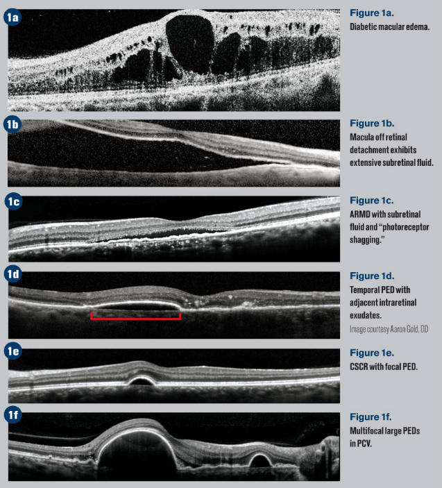
Over the last decade, there has been rapid growth in the management of glaucoma. Innovative imaging technologies that aid in the diagnosis of this vision-threatening disease have also been on the rise. Instead of relying on a single diagnostic tool, clinicians may consider leveraging the benefits of several modalities available today.
Haag-Streit Surgical recently announced it added several new features to its intraoperative OCT system, iOCT.
Heidelberg Engineering GmbH recently launched two new products, OCT2, the next generation OCT module for Spectralis diagnostic imaging, and the Heyex picture archiving and communication system (PACS).
Optovue recently launched its software-assisted OCT device, the iScan, in the United States.
Gulden Ophthalmics recently introduced Rowe OCT Model Eye, a solid-state tissue phantom that is used by clamping it to an instrument and initiating an OCT test.
A 56 year-old male patient came into the clinic recently for a second opinion concerning a previous injury to his right eye. A high-powered commercial laser in a physics laboratory was the source of the injury in 2005.
A mid-50s female was sent for imaging of a choroidal nevus that had been followed clinically for several years without observation of changes. There was no suspicion of growth or significant elevation. The purpose of the imaging studies was to establish digital baseline characteristics and to add additional documentation.
A 51 year-old female presented with recent-onset “distorted” vision in the right eye. The duration was described as 3 days and specifically, she used the terms “fragmented” and “pulsing” to describe her symptoms.
Optovue recently launched Avanit Widefield Enface OCT, an all-in-one solution for posterior and anterior high-speed, high-resolution OCT imaging, offering 70,000 A-scans per scond, Widefield 3D motion correction, and 3 micron digital resolution.
Katherine M. Mastrota, OD, shares pearls from “Reading Between the Lines: CT Interpretation” lecture at the 90th annual SECO International meeting.