
|Articles|February 17, 2022
- February digital edition 2022
- Volume 14
- Issue 2
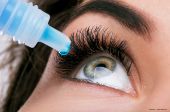
Dry eye disease onset at a younger age
Author(s)Scott Hauswirth, OD, FAAO
Screen time affecting millennials’ eye health.
Advertisement
Traditionally,
Prevalence increasing in all age groups
A 2019 retrospective analysis of data from the Department of Defense’s Military Health System (MHS) identified a DED population from 9.7 million MHS beneficiaries using diagnostic codes indicative of DED and prescriptions for cyclosporine ophthalmic emulsion.1 The investigators calculated the overall DED prevalence (2005-2012) and incidence (2008-2012), stratifying patients by sex and age group (18-39, 40-49, and ≥ 50 years old).
Overall prevalence, annual prevalence, and incidence were found to increase over time for all demographics. The annual prevalence tripled from 2005 to 2012, the authors noted. Although the annual prevalence increased sharply among beneficiaries older than 50, it also rose steadily for younger age groups.
Another study examining meibomian gland anatomy via meibography by Preeya Gupta, MD, and colleagues looked at 99 patients aged 4 to 17 years and found that 42% had evidence of meibomian gland atrophy and 37% had any evidence of meibomian gland tortuosity.2
Despite the changes in anatomic structure commonly associated with dry eye in adults, Standard Patient Evaluation of Eye Dryness Questionnaire (SPEED) scores were not statistically different. What this tells us is that anatomic changes may actually precede symptoms and that we should be paying more attention to the signs and symptoms of DED in younger patents.
Although many experts believe that the increasing ocular surface disease (OSD) seen in very young patients is linked to the proliferation of increased screen time, we do not yet have enough data to tease this out with a high degree of certainty. Screen time, although not the only factor, likely plays a role in exacerbating disease.
Developmental factors, genetic predispositions, and other environmental factors may also contribute to altering meibomian gland architecture. For example, blepharitis and allergic or atopic conjunctivitis lead to higher incidences of meibomian gland alterations due to the associated inflammation that affects gland anatomy and function.3,4 Prevalence of allergic conjunctivitis has been increasing over the past several decades (Miyazaki D et al, Allergol Int. 2020) and this may also be contributing to increased incidence of MGD.5
More millennials with OSD
The Pew Research Center identifies millennials as individuals born between 1981 and 1996. This group comprises of 72.26 million Americans—a larger cohort than baby boomers.6 These younger patients are noticing increased symptoms of OSD, and they are presenting to our offices in higher numbers.
In my experience, this demographic seems to be hungrier for information, and explanation and education seem to go further —they are eager to learn about their conditions and how best to help themselves. Describing the factors and stressors that contribute to their symptoms helps them grasp the importance of the therapies recommended for them.
My approach is to explain that what they are experiencing is consistent with tear film dysfunction and DED. I say, “Let’s put the puzzle pieces together by taking a look at environment, your daily routine, and when your discomfort is most noticeable.”
Typically, this is an easy conversation as these patients recognize that their eyes generally feel worse at the end of the day after they have been on the computer for a long time.
For many patients, there is no compromise when it comes to screen time—they work on computers, tablets, and cell phones. We cannot eliminate these devices, so the alternative is to teach good habits that include taking frequent, timed breaks and include therapies such as conscious blinking, or utilizing reminders to initiate a slow full blink, as these devices have been linked to decreased blink rate and amplitude (Portello, 2013).7
Implementing these therapies can be a challenge, but this is where education helps. In general, these management strategies are well received by my patients. I make them aware that their engagement with computer activity or whatever screen they are working on creates a disruption in the normal blink frequency and amplitude and leads to higher evaporative stress. I counsel them to find natural cues (such as changes in browsers or turning to a different screen) to do conscious blinking consisting of 1 or 2 slow full blinks, for example, and make sure they stop and pause, taking breaks from their screens every 15 to 20 minutes.
Providing foundational education
With all groups, but especially for millennials, education on other contributing factors such as dietary choices and medications are also well-received. Since a significant percentage of this population take systemic medications such as antidepressants or antianxiety medications that can be contributors to dry eye, it is helpful to review the contributions that these medications can make. I do not tell patients that they need to stop taking these agents—rather, I let them know that eye dryness is an expected adverse effect, which we will do our best to mitigate.
Describing how diet and nutritional supplements such as HydroEye (ScienceBased Health) affect ocular health is also foundational. The primary component of HydroEye’s patented formulation is the fatty acid gamma linolenic acid (GLA) derived from black currant seed oil. This unique anti-inflammatory omega-6, not found at meaningful levels in our diet, can play an important role in modulating the inflammatory response.8
GLA is present in breast milk and is naturally produced in the body via conversion from other omegas. It is a precursor to the anti-inflammatory prostaglandin PGE1—which promotes tear production and fights inflammation—and has been validated in a variety of clinical trials for improving dry eye symptoms in a range of patient populations.9-15 The supplement includes a specific balance of other omegas to provide dry eye relief.
Particularly among this demographic, one of the main reasons I hear about a lack of adherence with warm compresses is regarding the value of time. Patients do not want to take 15 minutes out of their day to sit with their eyes closed. I often describe this to patients as “potential spa time,” but younger patients often want to stay engaged with their world without that interruption.
TearRestore has developed a mask with an open-eye concept, which allows people to continue to function and engage with their life while they are using it. I am currently running a clinical trial to see how the TearRestore mask affects the tear film after a single application and over the course of a month (NCT04309799).
Natural, homeopathic interventions may help patients avoid medications—a message that is well received in this demographic. We do not always jump right into writing a prescription—we start with the basics such as nutrition, lubrication, heat therapy, and hygiene, laying a foundation and seeing how patients do with those measures. We can always add additional therapies if appropriate.
An additional clue to identifying OSD in younger patients is contact lens intolerance or decreased tolerance to wear time. Although they may not show high levels of epitheliopathy and staining, contact lens wearers will note a significant decrease in their ability to wear their lenses comfortably all day. The 2 most important tests to perform are tear breakup time and diagnostic meibomian gland expression. Often these 2 metrics are correlated.
In it for the long haul
We are largely a “right now” society, and certainly millennials have that mind-set. There is rarely a single magic bullet that will take care of dry eye. Rather, it is a combination of strategies that is usually most beneficial. I explain to the patient that we will work together to get them feeling better, but it will take some effort on their part. The dental model involving at home and occasional in-office therapies that we often talk about with DED is very applicable to younger individuals, and it tells them that their ocular health has to be managed for the long haul and not just the short term.
References
1. Dana R, Bradley JL, Guerin A, et al. Estimated prevalence and incidence of dry eye disease based on coding analysis of a large, all-age United States health care system. Am J Ophthalmol. 2019;202:47-54. doi:10.1016/j.ajo.2019.01.026
2. Gupta PK, Stevens MN, Kashyap N, Priestley Y. Prevalence of meibomian gland atrophy in a pediatric population. Cornea. 2018;37(4):426-430. doi:10.1097/ICO.0000000000001476
3. Ibrahim OMA, Matsumoto Y, Dogru M, et al. In vivo confocal microscopy evaluation of meibomian gland dysfunction in atopic-keratoconjunctivitis patients. Ophthalmology. 2012;119(10):1961-1968. doi:10.1016/j.ophtha.2012.04.001
4. Wei Q, Le Q, Hong J, Xiang J, Wei A, Xu J. In vivo microscopy of meibomian glands and palpebral conjunctiva in vernal keratoconjunctivitis. Indian J Ophthalmol. 2015;63(4):327-330. doi:10.4103/0301-4738.158073
5. Miyazaki D, Fukagawa K, Okamoto S, et al. Epidemiological aspects of allergic conjunctivitis. Allergol Int. 2020 Oct;69(4):487-495. doi: 10.1016/j.alit.2020.06.004
6. Statista. Resident population in the United States in 2020, by generation. Accessed December 7, 2021. https://www.statista.com/statistics/797321/us-population-by-generation/
7. Portello JK, Rosenfield M, Chu CA. Blink rate, incomplete blinks and computer vision syndrome. Optom Vis Sci. 2013 May;90(5):482-7. doi: 10.1097/OPX.0b013e31828f09a7
8. Kapoor R, Huang YS. Gamma linolenic acid: an anti-inflammatory omega-6 fatty acid (review). Curr Pharm Biotech. 2006;7(6):531-534. doi:10.2174/138920106779116874
9. Barabino S, Rolando M, Camicione P, et al. Systemic linoleic and gamma-linolenic acid therapy in dry eye syndrome with an inflammatory component. Cornea. 2003;22(2):97-101. doi:10.1097/00003226-200303000-00002
10. Macrì A, Giuffrida S, Amico V, Lester M, Traverso CE. Effect of linoleic acid and gamma-linolenic acid on tear production, tear clearance and on the ocular surface after photorefractive keratectomy. Graefes Arch Clin Exp Ophthalmol. 2003;241(7):561-566. doi:10.1007/s00417-003-0685-x
11. Aragona P, Bucolo C, Spinella R, Giuffrida S, Ferreri G. Systemic omega-6 essential fatty acid treatment and pge1 tear content in Sjögren’s syndrome patients. Invest Ophthalmol Vis Sci. 2005;46(12):4474-4479. doi:10.1167/iovs.04-1394
12. Kokke KH, Morris JA, Lawrenson JG. Oral omega-6 essential fatty acid treatment in contact lens associated dry eye. Cont Lens Anterior Eye. 2008;31(3):141-146. doi:10.1016/j.clae.2007.12.001
13. Pinna A, Piccinini P, Carta F. Effect of oral linoleic and gamma-linolenic acid on meibomian gland dysfunction. Cornea. 2007;26(3):260-264. doi:10.1097/ICO.0b013e318033d79b
14. Brignole-Baudouin F, Baudouin C, Aragona P, et al. A multicentre, double-masked, randomized, controlled trial assessing the effect of oral supplementation of omega-3 and omega-6 fatty acids on a conjunctival inflammatory marker in dry eye patients. Acta Ophthalmol. 2011;89(7):e591-e597. doi:10.1111/j.1755-3768.2011.02196.x
15. Sheppard JD, Singh R, McClellan AJ, et al. Long-term supplementation with n-6 and n-3 PUFAs improves moderate-to-severe keratoconjunctivitis sicca: a randomized double-blind clinical trial. Cornea. 2013;32(10):1297-1304. doi:10.1097/ICO.0b013e318299549c
Articles in this issue
over 4 years ago
When lifestyle and nutrition failover 4 years ago
Study: EHR notes may enhance, prolong racial biasover 4 years ago
Heru advances visual field assessmentsover 4 years ago
These digital tools for optometric prescribing are eye openingover 4 years ago
Top digital resources for prescribingover 4 years ago
Better flow for better patient careover 4 years ago
Retinal considerations crucial for accurate biometryover 4 years ago
MIGS dramatically transforms glaucoma managementAdvertisement
Related Content
Advertisement

Latest CME


Advertisement
Advertisement
Trending on Optometry Times - Clinical News & Expert Optometrist Insights
1
AOA 2026: Patterns of response to treatment with lifitegrast ophthalmic solution
2
Semaglutide initiation and rapid DR progression: a case report
3
AOA 2026: What the recent Oasys parameter expansion means for the Acuvue lens family
4
AOA 2026: High myopia's consequences on the retina
5