
Publication|Articles|December 8, 2025
- November/December digital edition 2025
- Volume 17
- Issue 06
How to put wearable virtual reality technology to work in your practice
Author(s)Bradford R. Ripps, OD
Fact checked by: Sheryl Stevenson, Ron Panarotti
VR technology captures not only visual fields but a full “stack” of pretest results.
Advertisement
As an acknowledged techie, I bought a Meta Quest virtual reality (VR) headset when it was introduced and have enjoyed using it for gaming and entertainment. I was struck by the idea that virtual and augmented reality devices could play a role in clinical practice. Sure enough, they have been rapidly evolving in that direction.
There are now 6 commercially available, FDA-registered wearable VR devices with the ability to perform perimetry plus a wide variety of other ophthalmic tests, including ocular motility, cover testing, color vision, pupil response, and visual acuity. Several years ago, we began using a VR perimetry device (Heru Prime; Heru) developed at the Bascom Palmer Eye Institute in Miami, Florida, as a screening tool in my practice. Today, it is fully integrated into our pretest process and has replaced the Humphrey Field Analyzer (HFA; ZEISS) for all our automated perimetry.
Visual field testing
We routinely use the VR headset for suprathreshold visual field (VF) screening for all our patients 10 years or older. The device can also perform 24-2 or 10-2 full-threshold VF testing, Esterman VFs, or confrontation fields as needed for those with risk factors or diagnoses that warrant full-threshold testing. Initially, I thought we would still need conventional perimetry for our patients with glaucoma. As the VR device capabilities have improved and our confidence in the results has grown, we now use it to follow all our patients with glaucoma as well. The Heru headset that we use has been shown to be highly correlated with the HFA Swedish Interactive Threshold Algorithm Standard in both normal eyes and eyes with glaucoma.1
Traditional perimetry is well tested and standardized. However, perimetry devices are large, challenging, or impossible to use for patients with mobility issues or those who use wheelchairs. They are also time-consuming. Patient fatigue during testing or staff skill level can affect the accuracy of the results. With a VR headset, it is simple to incorporate screening VFs for more patients. Depending on the device, VF testing can be performed in as little as 4 to 8 minutes. It is not uncommon for a patient to come in for a routine vision examination with no complaints or medical diagnoses, and for a VF defect to be detected with the test. Recently, I saw a patient who had screening perimetry failure. When the patient returned to the office 2 days later for full-threshold perimetry, it confirmed loss of VF. He had experienced an unrecognized stroke that affected his vision.
Patient feedback has been overwhelmingly positive. Younger patients who might use gaming headsets think the VR headset is cool, and even our older patients have not had any difficulty using it. Patients find it comfortable to wear and use, and do not fall asleep during VF testing, as they sometimes did with conventional perimetry. Interestingly, patients do not feel like it is the same test as the perimetry testing to which they were accustomed. They tell us it feels like they are playing a game.
A full pretest stack
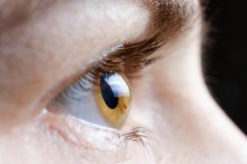
In our practice, patients check in and answer some history questions on a tablet computer and go directly to the pretest area. Typically, they start with the VR headset (Figure;
With the patient wearing the VR headset for only a few minutes, we are able to capture not only VFs but also a full “stack” of pretest results, ensuring that nothing is missed. Every patient undergoes Ishihara color vision testing, for example. Being able to generate documentation of test results for the patient chart is advantageous. Historically, I would make a notation of “normal” for cover testing, extraocular motility, and pupil response. Now I have a report generated by the headset with objective test data for the patient record, along with video playback that we can reference later or compare with future tests. A PDF of the results is attached to the patient’s electronic health record.
The quantitative pupillometry testing that is captured by the VR headset is impressive. There is significant evidence that changes in pupil response are among the earliest signs of glaucomatous and diabetic neuroretinal changes.2-5 Objective pupillometry quantifies pupillary light reflex dynamics affected by glaucoma; identifies abnormalities in pupil velocity, amplitude, and response latency; and evaluates intrinsically photosensitive retinal ganglion cells to provide early warning signs even before patients have lost VF. It provides much more data than manual pupillometry and a quick notation of “normally reactive pupils.” For those suspected of having glaucoma, in particular, I find VR pupillometry to be complementary to ultrawidefield imaging, providing a more complete picture of both anatomical and functional status.
A VR headset shifts many tasks from the physician to staff, freeing up my time in the examination room to interpret results and talk to the patient about their concerns or a new treatment plan. I love being able to see the testing results before I start refracting the patient.
Evaluating VR headsets
For those considering incorporating VR headset testing in the office, it is important to know that not all VR systems are equal when it comes to perimetry performance or other metrics. For example, some devices struggle with localized defects or are unreliable in moderate or advanced glaucoma, whereas others have demonstrated reliability across a broad dynamic range. Vera et al recently published a robust, systematic review of commercially available VR perimetry devices that have been compared with standard HFA perimetry in published studies.6 Of those studied, results were variable. The most extensively studied VR headset is the Olleyes VisuALL system, with 5 published studies, but it has been shown to have limited use in early disease detection, making it less suitable for glaucoma screening.6 Heru Prime offered the most consistent clinical validity and the closest correlation to all HFA measures, according to the review, but more published studies are needed.6
Integrating a new device into the clinic workflow always presents some challenges, but my staff and patients love using a VR headset for pretesting. New patients tell me they have not experienced an eye examination like the one in our office. To me, it reinforces the message I want to communicate: Patients can count on me to offer them the latest and greatest technology, whether that is in diagnostic technology or contact lenses. I believe we are at the beginning of the learning curve for what VR devices can deliver for the modern optometric practice and for our patients.
References
Johnson C, Sayed A, McSoley J, et al. Comparison of visual field test measurements with a novel approach on a wearable headset to standard automated perimetry. J Glaucoma. 2023;32(8):647-657. doi:10.1097/IJG.0000000000002238
Chang DS, Arora KS, Boland MV, Supakontanasan W, Friedman DS. Development and validation of an associative model for the detection of glaucoma using pupillography. Am J Ophthalmol. 2013;156(6):1285-1296.e2. doi:10.1016/j.ajo.2013.07.026
Adhikari P, Zele AJ, Thomas R, Feigl B. Quadrant field pupillometry detects melanopsin dysfunction in glaucoma suspects and early glaucoma. Sci Rep. 2016;6:33373. doi:10.1038/srep33373
Bayraktar S, Hondur G, Şekeroğlu MA, Şen E. Evaluation of static and dynamic pupillary functions in early-stage primary open angle glaucoma.
J Glaucoma. 2023;32(7):e90-e94. doi:10.1097/IJG.0000000000002212Feigl B, Zele AJ, Fader SM, et al. The post-illumination pupil response of melanopsin-expressing intrinsically photosensitive retinal ganglion cells in diabetes. Acta Ophthalmol. 2012;90(3):e230-e234. doi:10.1111/j.1755-3768.2011.02226.x
Vera J, Glazier AN, Dunbar MT, Ripkin D, Nafey M. Evaluating the clinical validity of commercially available virtual reality headsets for visual field testing: a systematic review. Vision (Basel). 2025;9(4):80. doi:10.3390/vision9040080
Articles in this issue
7 months ago
The A to Zzzs of ocular health7 months ago
The microbiome in diabetes and DRAdvertisement
Related Content
Advertisement


Latest CME


Advertisement
Advertisement
Trending on Optometry Times - Clinical News & Expert Optometrist Insights
1
Self-screening for ocular surface malignancies using a smartphone
2
Epion Therapeutics completes phase 3 trials of EpiSmart epithelium-on cross-linking for keratoconus
3
Johnson & Johnson invests $1B in ACUVUE contact lens manufacturing
4
Searching for Silence: Corneal Sensitivity Testing in NK
5