
Publication|Articles|August 11, 2025
- July/August digital edition 2025
- Volume 17
- Issue 04
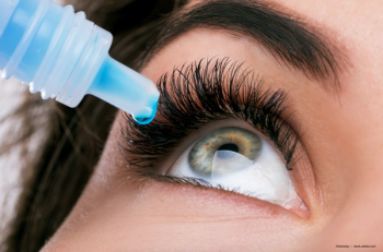
Grow your practice through anterior segment specialization
Author(s)Randy Charrier, OD, FAAO, Dipl CCLRT
In a climate of shrinking reimbursements and rising costs, finding your niche isn’t just smart—it’s essential.
Advertisement
My journey into anterior segment care began while leading the specialty contact lens clinic at the Naval Medical Center in San Diego, California. There, I treated complex corneal conditions and postsurgical complications in patients from around the globe. Later, I transitioned into private practice, taking over a specialty clinic from a retiring optometrist who had spent decades treating patients with corneal transplants or keratoconus.
Today, with mounting financial pressures on private practice optometry, many clinicians are searching for sustainable ways to maintain profitability and purpose. Specializing in anterior segment care has allowed me to build a distinctive brand, offer high-value services, and meet a critical need in my community while preserving revenue in an increasingly challenging environment.
Over the years, my clinic has become one of the leading providers of keratoconus care in the country. I’ve learned firsthand that developing a niche offers multiple advantages, such as the following:
- Stronger brand identity in a crowded market
- Ability to command premium pricing for expert care
- Increased referrals from general optometrists and ophthalmologists
- Higher patient retention due to trust and specialization
The power of advanced imaging
Advancements in imaging have significantly improved the precision of anterior segment disease detection. Technologies such as swept-source optical coherence tomography, corneal topography, and corneal tomography now allow for earlier and more accurate diagnosis of corneal irregularities such as keratoconus. These tools help clinicians map the shape, curvature, and biomechanical properties of the cornea with unprecedented precision, enhancing diagnostic accuracy.
Recently, I incorporated the Anterion multidisciplinary imaging platform by Heidelberg Engineering into my practice. This system captures ultra-high resolution, 3D images of the anterior segment, giving me the power to identify subtle degenerative changes, diagnose complex corneal conditions, and tailor treatment plans to each patient’s individual needs.
Early detection of keratoconus is critical: It opens the door to timely intervention with specialty scleral lenses or proactive treatments that may slow disease progression. Modern scleral lenses incorporate advanced technologies, including wavefront-guided and digitally molded designs. Pairing these innovations with high-quality diagnostics increases clinical efficiency and gives me confidence in the initial diagnosis and long-term monitoring.
Patients also respond positively to this technology. High-resolution imaging builds trust and reinforces the perception of precision and cutting-edge care. For many, it becomes a reason to stay with our clinic.
Investing in tech, elevating your brand
The ability to diagnose and manage complex cases with confidence has transformed my practice into a destination for advanced eye care. Tools that provide multidisciplinary diagnostics do more than improve clinical outcomes; they also differentiate the practice in a crowded market and create a pathway to sustained profitability.
These cases aren’t easy. Many of these patients have already seen multiple providers and carry a history of frustration and failed solutions. But therein lies the opportunity to make a real impact, change lives, and create loyal patients who become advocates for your expertise.
Anterior segment technology also enhances other areas of care. Orthokeratology, for instance, has become a widely embraced specialty within optometry. Imaging is key in tracking corneal changes and evaluating treatment success. For me, investing in a comprehensive platform that integrates topography, biometry, and epithelial mapping was the right decision not only for my practice, but for my patients. Having that full-picture view of each patient’s cornea has truly transformed my clinical approach.
Building expertise, expanding influence
For optometrists interested in anterior segment specialization, I encourage investing in ongoing education. Attend hands-on workshops, enroll in clinical courses, and seek mentorship opportunities from experienced specialists. Observing in high-level clinics and training with advanced instrumentation can dramatically accelerate your growth and confidence in managing complex cases.
You can also enhance your professional presence by contributing to academic journals, publishing case reports, and participating in relevant conferences. Staying connected to current research and cultivating peer networks opens doors to collaborative learning and innovative patient care. At my practice, we function as a teaching facility because we believe in investing in the future of our profession through education and mentorship.
Final thoughts
Optometrists who commit to anterior segment specialization and embrace state-of-the-art imaging technology can redefine their value to patients and referring providers. By creating a high-impact niche, you not only weather the pressures of declining reimbursements and rising costs, but you also position yourself as a leader in advanced vision care.
Articles in this issue
10 months ago
Myopia is an escalating global health crisis10 months ago
Inside genetics and glaucoma11 months ago
Embracing dry eye11 months ago
A case of orthokeratology for high myopiaAdvertisement
Related Content
Advertisement


Latest CME


Advertisement
Advertisement
Trending on Optometry Times - Clinical News & Expert Optometrist Insights
1
Johnson & Johnson invests $1B in ACUVUE contact lens manufacturing
2
Epion Therapeutics completes phase 3 trials of EpiSmart epithelium-on cross-linking for keratoconus
3
AOA 2026: Managing ocular toxicities in patients with cancer
4
Saptalis launches cyclosporine emulsion 0.05% for dry eye
5