Patient Case #1: Managing Dry Eye Before Cataract Surgery
Selina McGee, OD, FAAO, presents a patient case referred for ocular surface optimization in preparation for cataract surgery.
Selina McGee, OD, FAAO: Hello, I'm Dr Selena McGee. Thank you for joining us. I am the visionary founder and chief optometrist at Bespoke Vision in Edmond, Oklahoma. And I'm excited to share 2 patient cases with you as we discussed recently at our Ophthalmology Times® roundtable.
Our first case is going to be a 78-year-old [man]. He came into my clinic with difficulty adjusting to different light levels for the past 6 months, meaning that he would complain that as he would come inside, it would take a bit of time for him to adjust, and then when he would go outside as well. And then he was starting to struggle a little bit with just having good-quality vision with his mono-vision contact lens and his left eye that he had worn for many years.
He's retired, but he is an avid hobby photographer. So he travels all over the world doing these pretty extensive and in-depth trips on photography tours. [He’s a] very interesting patient with some detailed needs for his vision and how he utilizes his vision every day. When we walk through this case history, his review of systems is unremarkable. He does have an ocular history of mild macular drusen in both eyes. He is currently taking an AREDS2 medication. And when we also look at his entrance testing, his visual acuity is 20/30 right eye pinholes to 20/20 minus a few letters. His left eye is 20/200, but remember, this is his mono-vision eye. He does pinhole to 20/25. And with both eyes working together, he’s 20/25 at distance minus and then near 20/20 minus. But those are all soft numbers. So that's an important distinction when we look at visual acuity: [Although patients] measure well on the Snellen chart doesn't necessarily mean they have high-quality vision.
When you look at his refraction, his right eye was Plano, but I couldn't get him any better even behind the phoropter to 20/25, even though he pinholed a little better. And then his left eye is a –1.50 with a little bit of astigmatism. And we were able to get him to 20/20 on that side. His reading ADD is 2.25. And we were able to get him 20/20 in the lane. So PERRLA [pupils, equal, round, reactive to, light, accommodation] is normal, EOM [extraocular movement] is normal. Visual field, unremarkable. But I do a speed questionnaire on every one of my comprehensive exam patients because I have the mindset that when I see a patient, they have dry eye until they prove that they don't.
And I do that with [patients with] glaucoma as well. Just like when we do IOP [intraocular pressure], and we look at optic nerve and we look at ganglion cell complex and visual fields, those are all things to ensure that the patient sitting in front of us doesn't have glaucoma. And so I have the same type of testing for dry eye so that I ensure the patient does not have dry eye. And one of those is utilizing a speed questionnaire. He was 8 out of 28, which is significant. Anything in my clinic over 6, we have standing orders to perform osmolarity and MMP9 testing to look for inflammation. So his osmolarity was 314 and 310, which is high. And remember that with osmolarity, anything above 308 is significant. And anything that has a difference between each eye of greater than 8 is also significant. So he was significant symptomatically.
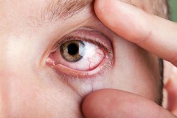
When we look at inflammation, he had right eye positive, left eye negative. And then we look at IOP was 14 and 15, and we did that with eye care. As we continue through the ocular exam, his anterior segment showed collarettes, which is pathognomonic for Demodex blepharitis. To ascertain that, I always have patients look down. [Next,] I'm lifting the skin so that I can make sure that I see the base of the lashes. And I'm doing that for several reasons. One, I'm looking for collarettes. Two, I'm looking for staph blepharitis.
I'm also looking for telangiectatic vessels that are right at that lid margin. Some patients will have lash extensions, other things that I want to be able to address and look at and make sure that things are healthy along the eyelid margin. An important component is having the patient look down. His bulbar and palpebral conjunctiva are white and quiet. The third piece, when I look at patients to make sure they don't have dry eye issues, I use a speed questionnaire. We have to look at corneal staining. And the other one is pushing on the meibomian glands and looking to see if they are functional. I will push on glands, temporal, medial, and then central to see they are functioning and what's the consistency of the meibum.
[This patient] was actually 0 out of 5 in all 3 of those areas, so completely nonfunctional. And then his corneal epithelium showed inferior SPK [superficial punctate keratitis], and his tear break-up time was lower than 10, at 8 and 6. He did have a normal tear meniscus height at 0.2 and 0.25. I have this measured with tear meniscus height, but you don't have to have that measured. You can look at a patient with their sodium fluorescein staining and be able to see what's abnormal.
Don't think that you have to have really sophisticated equipment to be able to do all of those things. You can do that simply with just vital dye. His anterior chamber was deep and quiet, the iris was normal. He did have +2 to 3 NS cataracts, OU. [Also, his] posterior, his C/D was normal at 0.2. And he does have macular drusen that we already knew about, but he also has a mild epiretinal membrane. We see that with the Optos and his OCT [optical coherence tomography] photos. The vessels were all normal. So when we think about our patients and needing to optimize the ocular surface and when we're thinking about cataract surgery, we absolutely have to optimize the ocular surface before we properly assess the patient and get their measurements for cataract surgery so that they have the best possible outcome.
When we talk to patients, we also want to talk about what's available with lenses now because we want to marry the technology we have currently in eye care to the patient's needs and desires.
He does desire the least dependence on spectacles and contact lenses. This is a [man who] was in Antarctica and keeping up with glasses and contact lenses in that kind of environment is not much fun. [It’s very] important that we understand our patients and we as optometrists have a longstanding relationship typically with our patients. We can really help our surgical colleagues by having these conversations early, having the ocular surface optimized. When we send our patients to the ophthalmologist for cataract surgery, we get great measurements so that we have good information coming in. The patient has the best-case scenario and the best possibility of having the desired outcome that…the surgeon, the optometrist, and the patient desires.
Transcript is AI-generated and edited for clarity and readability.
Newsletter
Want more insights like this? Subscribe to Optometry Times and get clinical pearls and practice tips delivered straight to your inbox.


