- January digital edition 2023
- Volume 15
- Issue 01
Reviewing a patient’s systemic history is critical when treating for OSD
Staying up-to-date on all things ocular is imperative in ocular surface disease.
When triaging patients suspected of having ocular surface disease, reviewing systemic history and medications is critical. As the Tear Film and Ocular Surface Society Dry Eye Workshop II reported, there are myriad systemic medications associated with dry eye, and the list grows every year1 as more adverse effects (AEs) from old medications are reported and new medications are brought to market. Below is a unique case of conjunctivitis with limbitis in a patient on dupilumab (Dupixent; Sanofi and Regeneron Pharmaceuticals), a systemic medication used in the treatment of atopic dermatitis that has been associated with a variety of AEs involving the anterior eye.
Case report
A male aged 39 years with a history of atopic dermatitis, chronic allergic conjunctivitis, and periorbital eczema presented to the clinic with a complaint of a painless, red right eye with tearing and discharge of mucus for 1 month. Review of systemic medical history revealed metabolic syndrome, type 2 diabetes, and hypertension, for which he was medicated with lisinopril, metformin, empagliflozin, and amlodipine. The patient had been receiving dupilumab injections for 14 months.
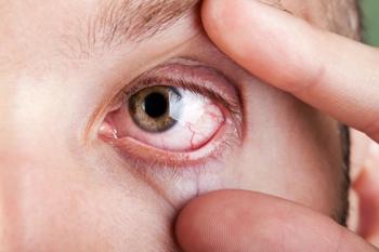
Vision was correctable to 20/20 and external testing was unremarkable. Intraocular pressure was normotensive. In external examination, Dennie lines (long-standing) would be appreciated (Figure 1A, Figure 1B). Slit lamp biomicroscopy of the right eye revealed 3+ bulbar conjunctival injection, significant limbal hyperemia and chemosis, and 3+ papillary reaction of the upper and lower palpebral conjunctiva.
The conjunctival injection blanched with 2.5% phenylephrine ophthalmic solution. There was scant mucus discharge and debris in the tear film. The cornea was clear with no staining with sodium fluorescein, although there was tear pooling adjacent to the areas of limbal hyperemia and a poorly wetting ocular surface (Figure 2). The anterior chamber was deep and quiet. Examination of the left eye showed 1+ papillary reaction of the superior and inferior palpebral conjunctiva and trace punctate epithelial erosions of the cornea. The dilated fundus examination was unremarkable in both eyes.
Due to the patient’s history of dupilumab use, chronicity of complaints, and absence of other signs or history indicating an infectious etiology, suspicion was high for a dupilumab-associated ocular AE. A panel of blood work was ordered to rule out an underlying autoimmune etiology for due diligence (all results returned negative). Prednisolone acetate 1% was initiated in the right eye 4 times daily.
At the 2-week follow-up, the patient’s symptoms and clinical signs were improved. Slit lamp biomicroscopy showed resolved limbal hyperemia, trace chemosis, and bulbar conjunctival injection in the affected eye. The cornea was clear, and the anterior chamber remained deep and quiet. With the improvement in clinical signs and symptoms, prednisolone acetate was tapered to 3 times daily for 2 weeks, then twice a day for 4 weeks (until the patient’s next examination).
Six weeks later, the patient presented to his follow-up examination with complete resolution of signs and symptoms in the right eye, but with new redness in the left eye that was identical in presentation to the right eye 2 months earlier. Prednisolone acetate 1% was initiated in the left eye 4 times daily, and the right eye was tapered to once a day. Lifitegrast ophthalmic solution 5% (Xiidra; Novartis) was also initiated twice a day in both eyes for long-term management. Two weeks later, the right eye remained quiet, and the left eye resolved. Prednisolone acetate 1% was tapered in the left eye.
Dupilumab conjunctivitis
Dupilumab is a subcutaneously injected monoclonal antibody that inhibits IL-4 and IL-13 signaling. It is FDA approved for the treatment of moderate-to-severe atopic dermatitis, asthma, eosinophilic esophagitis, and chronic rhinosinusitis with nasal polyps. Commonly reported AEs from dupilumab clinical trials include blepharitis and keratoconjunctivitis.2 Incidence of conjunctivitis in patients enrolled in clinical trials for the treatment of atopic dermatitis ranged from 8.6% to 21.44%.3-5
Recently, more severe reactions have been reported, including punctal stenosis, ectropion, limbal stem cell deficiency, ulcerative keratitis, and corneal perforation.6-10
Risk factors for dupilumab-associated conjunctivitis include greater severity of atopic dermatitis prior to treatment and history of conjunctivitis.11 Dupilumab does not appear associated with conjunctivitis in patients being treated for asthma, chronic rhinosinusitis with nasal polyps, or eosinophilic esophagitis.2 Interestingly, our patient had greater severity of atopic dermatitis in the right eye than in the left, represented by more severe Dennie lines in the right eye. This is also the eye that first presented with limbitis and conjunctivitis.
Management of dupilumab-associated ocular inflammation is dependent on the severity of the disease. Mild conjunctivitis can be managed conservatively with artificial tears, warm compresses, or antihistamine drops. Moderate-to-severe disease may necessitate anti-inflammatory topical ophthalmic drops or ointments such as corticosteroids or calcineurin inhibitors.11 Patients with punctal stenosis or cicatrizing conjunctivitis may benefit from surgical intervention if discontinuation of dupilumab and topical medication do not lead to resolution.6,7 Patients with corneal ulceration should be carefully monitored and referred to a corneal specialist if not responding to medical therapy and perforation is impending.8 Infectious etiology should be ruled out based on history, symptomology, clinical presentation, and culturing if necessary before initiating corticosteroid therapy.12 Long-term management with tacrolimus, cyclosporine, and lifitegrast have been shown to reduce risk of conjunctivitis recurrence.8,13
Ocular AEs are not an indication for discontinuation of dupilumab, although severe AEs may warrant discontinuation. During clinical trials, 80% of cases resolved without recurrence.2 Pretreatment of any preexisting ocular surface disease (eg, dry eye disease or blepharitis) may be beneficial to reduce the risk of dupilumab-associated conjunctivitis, and screening patients before initiating therapy may be prudent.10 Several pathophysiological mechanisms have been proposed for dupilumab-associated conjunctivitis, including an increase in Demodex mites as a result of altered cytokine activity, as well as IL-13–mediated decrease in mucus production and goblet cells. These may explain the benefits of a healthy ocular surface in preventing dupilumab-associated conjunctivitis.2
Conclusion
In conclusion, in cases of conjunctivitis with atypical presentation, a careful review of systems and medication history should occur. With dupilumab-associated intraocular inflammation specifically, it is important to rule out autoimmune-mediated ocular inflammation and infectious etiologies with appropriate serology and culturing as warranted, given the broad differential diagnosis.
As primary eye care providers, we interact regularly with patients of a wide variety of clinical backgrounds. We are often the only eye care providers a patient sees, making it important to stay up-to-date with ocular AE profiles of medications, new and old.
Dupilumab is an effective medication for atopic dermatitis that will be used with increasing prevalence. It recently gained approval for use in children with moderate-to-severe atopic dermatitis.14 It is important for the optometrist to identify and treat this manageable AE so that patients may continue to be treated with dupilumab for their chronic illness.
References
1. Gomes JAP, Azar DT, Baudouin C, et al. TFOS DEWS II iatrogenic report. Ocul Surf. 2017;15(3):511-538. doi:10.1016/j.jtos.2017.05.004
2. Akinlade B, Guttman-Yassky E, de Bruin-Weller M, et al. Conjunctivitis in dupilumab clinical trials. Br J Dermatol. 2019;181(3):459-473. doi:10.1111/bjd.17869
3. Simpson EL, Bieber T, Guttman-Yassky E, et al; SOLO 1 and SOLO 2 Investigators. Two phase 3 trials of dupilumab versus placebo in atopic dermatitis. N Engl J Med. 2016;375(24):2335-2348. doi:10.1056/NEJMoa1610020
4. Blauvelt A, de Bruin-Weller M, Gooderham M, et al. Long-term management of moderate-to-severe atopic dermatitis with dupilumab and concomitant topical corticosteroids (LIBERTY AD CHRONOS): a 1-year, randomised, double-blinded, placebo-controlled, phase 3 trial. Lancet. 2017;389(10086):2287-2303. doi:10.1016/S0140-6736(17)31191-1
5. de Bruin-Weller M, Thaçi D, Smith CH, et al. Dupilumab with concomitant topical corticosteroid treatment in adults with atopic dermatitis with an inadequate response or intolerance to ciclosporin A or when this treatment is medically inadvisable: a placebo-controlled, randomized phase III clinical trial (LIBERTY AD CAFÉ). Br J Dermatol. 2018;178(5):1083-1101. doi:10.1111/bjd.16156
6. Lee DH, Cohen LM, Yoon MK, Tao JP. Punctal stenosis associated with dupilumab therapy for atopic dermatitis. J Dermatolog Treat. 2021;32(7):737-740. doi:10.1080/09546634.2019.1711010
7. Nettis E, Guerriero S, Masciopinto L, Di Leo E, Macchia L. Dupilumab-induced bilateral cicatricial ectropion in real life. J Allergy Clin Immunol Pract. 2020;8(2):728-729. doi:10.1016/j.jaip.2019.10.015
8. Mehta U, Farid M. Dupilumab induced limbal stem cell deficiency. Int Med Case Rep J. 2021;14:275-278. doi:10.2147/IMCRJ.S308583
9. Li G, Berkenstock M, Soiberman U. Corneal ulceration associated with dupilumab use in a patient with atopic dermatitis. Am J Ophthalmol Case Rep. 2020;19:100848. doi:10.1016/j.ajoc.2020.100848
10. Phylactou M, Jabbour S, Ahmad S, Vasquez-Perez A. Corneal perforation in patients under treatment with dupilumab for atopic dermatitis. Cornea. 2022;41(8):981-985. doi:10.1097/ICO.0000000000002854
11. Agnihotri G, Shi K, Lio PA. A clinician’s guide to the recognition and management of dupilumab-associated conjunctivitis. Drugs R D. 2019;19(4):311-318. doi:10.1007/s40268-019-00288-x
12. Gooderham M, McDonald J, Papp K. Diagnosis and management of conjunctivitis for the dermatologist. J Cutan Med Surg. 2018;22(2):200-206. doi:10.1177/1203475417743233
13. Zirwas MJ, Wulff K, Beckman K. Lifitegrast add-on treatment for dupilumab-induced ocular surface disease (DIOSD): a novel case report. JAAD Case Rep. 2018;5(1):34-36. doi:10.1016/j.jdcr.2018.10.016
14. Napolitano M, Fabbrocini G, Neri I, et al. Dupilumab Treatment in Children Aged 6-11 Years With Atopic Dermatitis: A Multicentre, Real-Life Study. Paediatr Drugs. 2022;24(6):671-678. doi:10.1007/s40272-022-00531-0
Articles in this issue
about 3 years ago
Contact lenses can help patients with low visionabout 3 years ago
Surprising findings from the DRCR Retina Network Protocol AAabout 3 years ago
EVO ICL may be best-kept secret in refractive surgeryabout 3 years ago
Managing low vision in an aging populationabout 3 years ago
Patients stick with us, for better or for worseabout 3 years ago
Vision care includes dry eye careNewsletter
Want more insights like this? Subscribe to Optometry Times and get clinical pearls and practice tips delivered straight to your inbox.


