


Proper screenings, treatment, management needed for this increasingly prevalent disease.
Proper screenings, treatment, management needed for this increasingly prevalent disease.
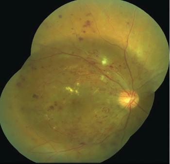
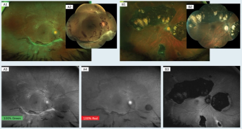
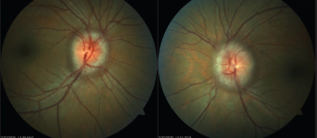
The patient’s ocular history was significant for severe nonproliferative diabetic retinopathy (NPDR) in the right eye (OD) and mild NPDR in the left eye (OS), which had been diagnosed 4 years previously at his last eye exam.

Korean investigators have found intermittent fasting may actually lead to a lower risk of developing AMD in the elderly population.

If approved, pegcetacoplan—an investigational, targeted C3 therapy—will be the first-ever treatment for GA.
Advancements enable better diagnosis, treatment of patients.

Presented during the 2022 ASRS meeting, results find that over 60% of participants could be treated every 4 months at 2 years—an increase from 45% at year 1.

A decade after the conclusion of the NIH-funded AREDS2 study, researchers found that the AREDS2 formula also reduces the risk of lung cancer.

Trial results demonstrated statistically significant improvement in the prespecified primary endpoint in BCVA at 13 months in the PBM treatment group over the sham-treatment group.
A. Paul Chous, OD, discusses his presentation, “Current treatment of diabetes" during the AOA's Optometry Meeting.
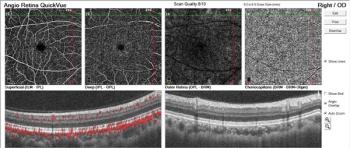
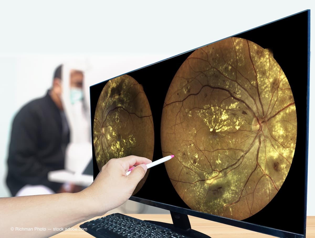
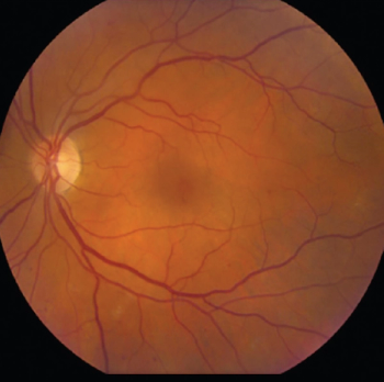
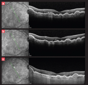
Case report details diagnosis based on imaging diagnostics.

Embracing this new model of care can allow optometric practices to flourish—and may enable earlier detection of conversion from intermediate dry to wet AMD.

Early diagnosis and intervention are crucial when managing this patient base.

All data were collected in the Fight Retinal Blindness! Registry.

This is the second FDA-approved indication for BEOVU; it was initially approved in 2019 for the treatment of wet AMD.

A decision by the FDA is expected in August 2022.
The ADA now recommends an individualized approach at diagnosis of diabetes as well as for adults who do not have symptoms to be screened at age 35 years.
Four patients present with previously undiagnosed conditions.

The company, formerly known as Clover Therapeutics, intends to use the funding to advance two lead drug candidates that target molecularly-defined AMD patient subtypes.

The company's technology allows for ECPs to detect, monitor, and treat AMD 3 years before clinical diagnosis.
A look at the latest developments in retinal disease, including clinical trials and advanced therapies.
Oxurion NV finds insufficient evidence of efficacy on key clinical endpoints for THR-687 in Part A of the INTEGRAL phase 2 trial; focus will now turn to an alternative candidate and trial.

New detailed, longer-term findings were presented at the 2022 ARVO meeting.
Diagnosis highlights overlooked treatments for this disease

Investigators report a correlation between the risk of 5-letter visual acuity (VA) loss at 24 months for eyes with clinically significant diabetic macular edema (CSDME) and good VA initially treated and eyes that were initially observed in routine clinical practice.

Gifted by philanthropist James Grosfeld, the money will fund the launching of a pioneering research initiative as one of the largest investments of resources in the United States.